Primary immunodeficiencies (PID) Clinical Update
ASCIA PID Clinical Update 2025

The content of this document is consistent with ASCIA primary immunodeficiencies (PID) e-training for health professionals.

The main purpose of this document and the ASCIA PID e-training course is to:

  • Increase awareness of PID assist health professionals, particularly general practitioners, paediatricians and general physicians.
  • Promote early recognition and referral to a clinical immunologist to improve the management and quality of life for patients with PID in Australia and New Zealand.

pdfASCIA HP Clinical Update PID 2025669.58 KB

Contents include:

1. Immunodeficiency overview

2. Recognition, diagnosis and management of PID

3. Immunoglobulin Replacement Therapy (IRT)

4. Severe Combined Immunodeficiency (SCID)

5. Antibody deficiency disorders

6. Complement disorders including HAE

7. Other PID syndromes

8. References and further information (See PDF for references)


1. Immunodeficiency overview

How does the immune system work?

The immune system is a collection of tissues (e.g. bone marrow, thymus, lymph nodes and spleen), cells (e.g. lymphocytes, neutrophils and macrophages) and molecules that can be broadly divided into two parts, the innate and adaptive immunity (Marshall et al., 2018). There can be significant overlap between these 2 systems.

Innate immune responses:

  • Are present before there is contact with pathogens.
  • Are limited in specificity.
  • Have no memory or long lived protection.
  • Are mediated by cells (e.g. neutrophils, natural killer [NK] cells) and circulating molecules (e.g. complement proteins).

Adaptive immune responses:

  • Are slower to initiate.
  • Respond specifically to antigens.
  • Result in immunological memory.
  • Are mediated by cells (T cells, B cells, dendritic cells) and circulating molecules (antibodies).

Effects of age on the immune system

Whilst newborns have maternal antibodies that crossed the placenta during pregnancy, their adaptive immune systems are still developing. Serum immunoglobulin (Ig) levels normally drop as maternal antibodies wane, before recovering as the newborn’s immune system develops (Palmeira et al., 2012).

With advanced age the immune system weakens, particularly the adaptive system, resulting in less efficient responses by T cells and less efficient B cells with less diversity in the immune system (Valiathan et al., 2016). This makes older patients more prone to infection and non-infectious diseases, such as cancer.

What are immunodeficiencies?

Immunodeficiencies are a heterogeneous group of disorders in which there is a defect in the normal function of the immune system (Raje and Dinakar, 2015).  Delayed recognition and diagnosis of immunodeficiencies leads to poor health outcomes and potentially premature death.

Infections are the predominant feature of immunodeficiency, particularly infections that may:

  • Be unusually persistent, severe, recurrent or resistant to treatment.
  • Involve unusual organisms or organisms of usually low virulence.
  • Involve body sites not usually prone to infection or multiple organs.

Immunodeficiencies may present at any age and are associated with an increased risk of certain types of autoimmune conditions and malignancies.

There are two types of immunodeficiencies:

  • Primary Immunodeficiencies (PID) - There are more than 550 different PID and these are mostly caused by inherited defects of the immune system (Bousfiha et al., 2025). This document focuses on PID.
  • Secondary Immunodeficiencies (SID) - These are usually caused as part of another disease or illness (e.g. severe burns, haematological malignancy), as a consequence of certain medications (e.g. chemotherapy) and some infections (e.g. HIV). SIDs are more common than PID and lead to an increased incidence of infection, malignancy or autoimmune disease, similar to PID (Ballow et al., 2022). 

Primary immunodeficiencies (PID) overview

PID are inherited defects that affect development and/or function of the immune system and result in increased susceptibility to infection, autoimmunity and malignancy.

More than 550 different immunodeficiency syndromes are now described and whilst individually PID are rare, collectively they make up a significant chronic disease. The prevalence of PID is estimated to be 1 in 1200 but is variable depending on specific disorder (e.g. IgA deficiency occurs in 1 in 500 whilst other PID occur in 1 in 10,000 – 1:1,000,000 people) (Lee and Gray, 2014; McCusker and Warrington, 2011).

More severe forms of PID typically become apparent in early life, such as severe combined immunodeficiency (SCID), with only a short asymptomatic period after birth (Dvorak et al., 2023). Early recognition of severe PID is critical as successful treatment outcomes are dependent on early diagnosis.

PID may present at a later age, with some PID such as common variable immunodeficiency (CVID) more commonly diagnosed in adults (Tam and Routes, 2013).

Despite major advances in molecular characterisation over the last 20 years, many patients remain undiagnosed or are diagnosed late, with adverse effects on morbidity and mortality. Treatment options for PID include prophylactic antibiotics, immunoglobulin replacement therapy (IRT) and in some PID, haematopoietic stem cell transplantation (HSCT), such as bone marrow transplantation (BMT) (Segundo and Condino-Neto, 2021). 

PID and the immune system

Although PID vary in many ways, they all result from a defect in one or more elements of immune system regulation or function, as shown in the following table.

Classification

Immune defect

Examples

Combined immunodeficiencies

T cells and B cells

Severe combined immunodeficiencies (SCID) e.g. X-linked SCID, RAG 1 and 2 deficiency

Combined immunodeficiencies with associated or syndromic features

T cells and B cells with other features

Ataxia-telangiectasia, DiGeorge syndrome,

Wiskott-Aldrich syndrome (WAS), Hyper-IgE syndromes (HIES), DOCK-8, NEMO

Predominantly antibody immunodeficiencies

B cells or plasma cells

X-linked agammaglobulinaemia, common variable immunodeficiency (CVID), specific antibody deficiency

Diseases of immune dysregulation

Susceptibility to haemophagocytosis or defects in regulation of immune system

Chediak-Higashi syndrome, familial haemophagocytic lymphohistiocytosis (HLH, FLH), X-linked lymphoproliferative syndrome

Congenital defects of phagocytes

Phagocytes number or function

Severe congenital neutropaenia, leukocyte adhesion deficiency, chronic granulomatous disease (CGD)

Defects of innate immunity

Innate immune system

IRAK-4 and MyD88 deficiencies, WHIM syndrome, TLR3 and UNC93B1 deficiencies, chronic mucocutaneous candidiasis

Autoinflammatory disorders

Cytokine overproduction

Familial Mediterranean fever, Hyper-IgD syndrome,

Muckle-Wells syndrome, familial cold autoinflammatory syndrome

Complement immunodeficiencies

Complement components

Deficiencies of components of the classic or alternate complement pathway (e.g. C5 or C6 deficiency),

C1 inhibitor deficiency / hereditary angioedema (HAE)

Phenocopies of PID

Arise from acquired mechanisms

(a)Phenocopies associated with somatic mutations e.g. autoimmune lymphoproliferative syndrome (ALPS-SFAG)

(b) Phenocopies associated with autoantibodies

e.g. chronic mucocutaneous candidiasis due to IL-17 autoantibody, adult-onset immunodeficiency due to INF-gamma autoantibody, pulmonary alveolar proteinosis due to Granulocyte-macrophage colony-stimulating factor (GM-CSF) autoantibody

Raje and Dinakar (2016)

2. Recognition, diagnosis and management of PID

There are seven early warning signs of PID (McCusker et al., 2018):

  1. An unusually large number of infections requiring treatment including:
    • Frequent middle ear infections (>8 per year).
    • More than one serious sinus infection per year.
    • More than one pneumonia infection per year.
    • Chronic suppurative sinus or lung disease in adults.
  1. Infections caused by unusual or opportunistic types of organisms including:
    • Organisms of low pathogenicity.
  1. Infections in unusual places including:
    • Perianal abscesses
    • Organ abscesses
  1. Infections that do not respond to treatment as normally expected, including:
    • Infections refractory to antibiotic treatment.
    • Rapid recurrence of infections after ceasing antibiotic treatment.
    • Persistent unexplained oral thrush after one year of age.
    • Severe infections (e.g. meningitis, osteomyelitis, pneumonia) requiring IV antibiotics.
    • Middle ear infections asssociated with a persistent purulent discharge.
    • Recurrent serious sinus infections.
    • Persistent production of purulent sputum.
  1. A child that does not grow or put on weight as expected, or weight loss in adults.
  1. A family history of immunodeficiency or abnormal infections, particularly if there is:
    • Shared kinship of parents.
    • A family history of fatal infective complications or unexplained infant deaths.
  1. Any other unusual symptoms related to infections, including:
    • Persistent diarrhoea.
    • Unusual infection related sequelae or syndromes.

Other features of PID (these mainly occur in adult patients with CVID) include :

      • Autoimmune cytopenias.
      • Granuloma formation at any site (often misdiagnosed as sarcoidosis).
      • Chronic enteropathy unresponsive to gluten withdrawal from diet.
      • Unexplained hepatomegaly especially in adults.
      • Unexplained splenomegaly in children or adults.
      • Lymphoid interstitial pneumonitis.

Delay in diagnosis is common

Severe PID may be recognised in early childhood.  However, many PID are undiagnosed into adolescence or adulthood.  According to a survey by the Immue Deficiencies Foundation (IDF) in 2007 amongst 1,300 people with PID, the average time from symptom onset to diagnosis is 12.4 years (Condino-Neto and Espinosa-Rosales, 2018).

The role of GPs, paediatricians and physicians in identifiying and managing patients with PID

GPs, paediatricians and adult medicine physicians, particularly respiratory medicine physicians and gastroenterologists, have an important role in identifying and managing patients with PID, including (O’Keefe et al., 2016):

      • Recognition of early symptoms and signs of PID.
      • Appropriate investigation and interpretation of test results.
      • Appropriate and timely referral to a clinical immunologist.
      • Appropriate follow up care, in conjunction with a clinical immunologist, particularly assessment of growth and development in children.
      • Management of general health issues of patients with PID.

Expected frequency of infections in childhood

The burden of infectious disease is high in children, even in individuals with healthy immune systems.  In Australia normal healthy toddlers can have up to 4-8 minor infections each year.  The number of infections can be increased up to 12 if the child attends childcare, has older siblings or has parents who smoke.

PID should be considered in patients with recurrent infections (more than expected for age), severe infections or infection with unusual or opportunistic organisms.

Frequency of infections in general childhood population: 

Age (yrs)

Infections/yr

0-2

6

3-4

5

5-9

4

10-14

3

Gray et al. (2010)

Testing for PID

Initial evaluation always starts with a thorough clinical history and physical examination.

Prediction of specific PID on the basis of a particular organism is difficult.  However, identification of certain types of microbial organisms may help provide clues as to the underlying defect in the immune system, as shown in the following table. 

Infections associated with PID

PID

Infections

Organisms

Cellular (combined T and B cell) immunodeficiencies

Systemic viral infections, intracellular bacteria, fungal infections

Bacteria: Pyogenic bacteria

Viruses: All

Mycobacteria: Non-TB including Bacillus Calmette-Guerin (BCG)

Fungi: Candida, Aspergillus

Protozoa: Pneumocystis jiroveci pneumonia (PJP), cryptosporidium

Antibody (B cell) deficiencies

Upper and lower respiratory tract infections, GI tract infections

Bacteria: Streptococcus pneumonia (pneumococcus), Haemophilis influenzae, Moraxella catarrhalis, Campylobacter

Viruses: Enteroviruses

Protozoa: Giardia

Phagocytic immune defects

Respiratory tract, cellulitis, skin abscesses, liver abscesses

Bacteria: Staphylococcus aureus, Pseudomonas aeruginosa

Mycobacteria: Non-TB including BCG

Fungi: Candida, Aspergillus

Complement immunodeficiencies

Meningitis, systemic bacterial infections

Bacteria: Neisseria, Streptococci

Oliveira and Fleisher (2010)

Antibody (B cell) deficiencies

Antibody deficiencies are the most common type of immunodeficiency and are associated with decreased ability to produce protective antibodies against pathogens. They usually present from three months after birth, once maternal antibodies transferred across the placenta have gone (Ozdemir, 2021).

Patients with antibody or humoral deficiencies generally have more frequent and/or prolonged infections compared to healthy individuals. Typically these patients suffer recurrent or severe bacterial infections of ears, sinuses and lungs, particularly with encapsulated organisms (e.g.pneumococcus, meningococcus, haemophilus) (Oliveira and Fleisher, 2010).

Evaluation of antibody (B cell) deficiencies

  • Antibody function can be tested both quantitatively and qualitatively.
  • Quantitative serum immunoglobulin (Ig) tests are used to detect abnormal levels of the 3 major classes of Ig – IgG, IgA and IgM (Loh et al., 2013).
  • A low level of IgG is termed ‘hypogammaglobulinaemia’ and its presence should be confirmed on repeat testing. Any underlying secondary causes of hypogammaglobulinaemia should be considered before referral to a clinical immunologist (Anderson et al., 2022).
  • Cellular immune testing for B cells should be performed in consultation with a clinical immunologist.
  • Assessment of specific antibody responses (e.g. tetanus and pneumococcus) may be required, usually in consultation with a clinical immunologist, in less severe forms of hypogammaglobulinaemia or when the clinical history is suggestive of an antibody deficiency disorder (Thong et al., 2023). Specific antibodies need to be correlated with vaccine history and age of patient.

Patients with abnormal results or patients with normal results who are of ongoing clinical concern should be referred to a clinical immunologist.

Cellular (combined T and B cell) immunodeficiencies

Cellular immunodeficiencies are much less common than antibody immune defects and are often present with defects of B cells and other immune cells (Buckley, 2002). Therefore they are sometimes called combined immunodeficiencies. Primary T cell deficiencies present early in life, with failure to thrive, opportunistic infections, candida and chronic diarrhoea.

If a cellular immunodeficiency is suspected, live vaccines need to be withheld and patient should be referred to a clinical immunologist for an urgent consultation.

Evaluation of cellular (combined T and B cell) immunodeficiencies

Tier 1 (GPs, paediatricians)

  • Full blood count
  • Particularly in children, transient lymphopenia is common. Persistent or profound lymphopenia should be followed up.
  • T cells comprise two thirds of lymphocytes in blood, therefore decreased T cells are typically associated with lymphopenia on a full blood count but normal T cells does not rule out cellular immunodeficiency.
  • Quantitative serum Ig tests are used to assess humoral immunity.

Tier 2 (Clinical immunologists and/or paediatricians)

  • Flow cytometry for T cells, B cells and NK cells.
  • HIV testing should be performed to exclude HIV.
  • Additional testing may be indicated.

Lee and Gray (2014) 

Severe T cell immunodeficiencies such as SCID are medical emergencies and patients need to be urgently referred to a clinical immunologist for continuation of diagnosis and treatment. (Firatoglu et al., 2025)

Phagocyte immune defects

Phagocyte abnormalities are due to defects in neutrophils or monocytes/macrophages and are commonly present with recurrent pyogenic or fungal skin infections and/or abscesses (Mickey et al., 2024).

Evaluation of phagocyte immune defects

Tier 1 (GPs, paediatricians)

  • Full blood count with blood film can assess for neutropenia although multiple time points are needed to exclude conditions with fluctuating neutrophil counts.
  • Blood films may show disorders associated with abnormal granules inside phagocytes.

Tier 2 (clinical immunologists)

  • If neutrophil numbers are normal, nitroblue tetrazolium (NBT) and/or dihydrorhodamine (DHR) tests may be used to assess for function.
  • Additional testing may be indicated.

Mickey et al. (2024)

Complement immunodeficiencies

Complement immunodeficiencies may be due to deficiencies in any on the components of the 3 complement pathways (McMurray et al., 2024).. Clinical indications include bacterial infections (especially Niesseria), autoimmunity and recurrent angioedema.  

Evaluation of complement immunodeficiencies

Initial screening should include C3, C4, AH50 and CH50.  In patients with recurrent angioedema C4 and C1 esterase levels should be tested (Bonilla et al., 2015). Additional testing including individual components may be indicated in consultation with a clinical immunologist.

When should patients be referred to a clinical immunologist?

Patients should be referred to a clinical immunologist when (O’Keefe et al., 2016):

  • They have early warning signs of PID
  • Results of initial screening suggests PID
  • Results of initial screening are confusing and diagnosis is unclear
  • Results do not confirm PID but there remains a high clinical suspicion for PID

Additional testing includes specialised molecular and genetic tests, that may be needed to confirm a diagnosis.

General care of PID patients

Management of patients with PID requires a multidisciplinary team.

Issues include:

  • Monitoring growth and ensuring adequate nutrition in children (Karhan et al., 2022).
  • Optimising exercise, sleep and stress management.
  • Ensuring good general hygiene (e.g. hand washing, dental hygiene)
  • Providing information about patient support organisations:
  • AusPIPS - auspips.org.au
  • HAE (Hereditary Angioedema) Australasia – haeaustralasia.org.au
  • Immune Deficiencies Foundation Australia (IDFA) – idfa.org.au
  • Immune Deficiencies Foundation New Zealand (IDFNZ) – idfnz.org.au

Specific therapies for PID

The following therapies need to be individualised according to the type of PID (Segundo and Condino-Neto, 2021):

  • Prophylactic antibiotics, antifungals, antivirals.
  • Immunoglobulin replacement therapy (IRT).
  • Immune modulatory drugs.
  • Gamma interferon.
  • Haemotopoetic stem cell transplant (HSCT).
  • Bone marrow transplantation (BMT).
  • Gene therapy.
  • Others – granulocyte-colony stimulating factor (G-CSF), polyethylene glycol-conjugated adenosine deaminase (PEG-ADA).

Infections in PID patients

Infections should be investigated and treated aggressively. It is important to (Refaat et al., 2024):

  • Notify microbiology laboratory to consider atypical or opportunistic infections.
  • Treat patient with broad spectrum antibiotics until organism is identified.
  • Consider fungal, mycobacterial, viral and protozoan therapy, according to specific condition.

Serology is not useful if patient is unable to make antibodies or is on immunoglobulin replacement therapy (IRT).

Curative therapies

Haemotopoetic stem cell transplant (HSCT)

Potential sources of stem cells include bone marrow, peripheral blood or umbilical cord blood (Castagnoli et al., 2019).

Gene therapy

This therapy is still experimental and is therefore not routinely available. It involves the use of viral organisms to replace the missing genes and correct the abnormality (Houghton and Booth, 2021).

PID and vaccinations

If a PID is suspected or confirmed, live attenuated vaccines should not be routinely administered as they may cause disease in immunodeficient patients. Issues regarding immunisation of these patients should be discussed with a clinical immunologist (Sobh and Bonilla, 2016).

Potential live vaccines include (Shearer et al., 2014):

  • MMR
  • Varicella 
  • BCG (given to at risk populations).
  • Rotavirus 
  • Yellow fever.

Meningococcal, pneumococcal and Haemophilus Influenza vaccines are recommended for patients with complement immunodeficiencies.

Current immunisation guidelines (ATAGI, 2022):  https://immunisationhandbook.health.gov.au/

Vaccination information for patients on immunoglobulin replacement therapy (IRT): https://immunisationhandbook.health.gov.au/contents/vaccination-for-special-risk-groups/vaccination-for-people-who-have-recently-received-normal-human-immunoglobulin-and-other-blood-products  

3. Immunoglobulin replacement therapy in PID 

Immunoglobulin replacement therapy (IRT)

Replacement of serum immunoglobulin with human gamma globulin fraction is the standard treatment for most patients with hypogammaglobulinaemia and some with specific antibody deficiencies (Sriaroon and Ballow, 2015). The aim is to replace immunoglobulin to maintain normal IgG levels. The dose used is individualised for each patient.

IRT may be given as intravenous immunoglobulin (IVIg) or subcutaneous immunoglobulin (SCIg) and pharmacokinetics differ according to administration route (Mallick et al., 2025). There are multiple brands that may change from time to time and rates of administration vary for different products.

Immunoglobulin products

Immunoglobulin products are made from pooled plasma from many healthy human donors (Fernández-Cruz et al., 2009). Plasma donors and plasma pools are screened for hepatitis B, hepatitis C and HIV. Additional viral inactivation steps such as heat treatment, enzyme treatment, detergent treatment and nanofiltration are performed.

Despite differences in manufacturing, all IgG products contain 97-98% IgG specific antibodies against broad spectrum of bacterial and viral pathogens, with traces of IgM and IgA.

Long term benefits of immunoglobulin replacement therapy

Both intravenous immunoglobulin (IVIg) and subcutaneous immunoglobulin (SCIg) (Jolles et al., 2005):

  • Are effective at reducing infections and hospitalisations.
  • Preserve organ function and reduce long term damage from recurrent infections.
  • Are associated with significant benefits to patient quality of life.
  • Improve the lifespan of patients.

Intravenous Immunoglobulin (IVIg) replacement therapy

Intravenous immunoglobulin (IVIg) replacement therapy (Li and Mahmood, 2024):

  • Is usually administered three to four weekly in hospital.
  • Leads to a high peak of IgG after infusion.
  • Levels decrease rapidly over few days then slowly decrease over next few weeks.

The majority of side effects of IVIg are mild and self-limiting and include headache, fever, chills, nausea, fatigue or flu-like illness (Guo et al., 2018). The frequency of side effects of IVIg may be linked to the rate of infusion.

Serious adverse events are rare but include anaphylaxis, aseptic meningitis, renal impairment and  thrombosis.

Subcutaneous Immunoglobulin (SCIg) replacement therapy

Subcutaneous immunoglobulin (SCIg) requires frequent administration (one to three times per week) by patients or carers at home. SCIg involves slow diffusion of IgG from subcutaneous tissue and the small and more frequent doses are associated with more consistent IgG levels (Jolles et al., 2015). Whilst systemic reactions to SCIg are much less common than with IVIg, local injection site reactions (e.g. redness, itching, swelling, discomfort) are common but improve with time (Cinetto et al., 2021).

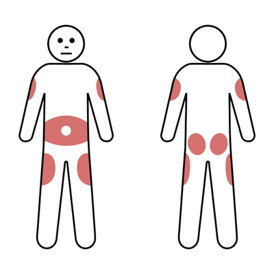
SCIg injections may be administered at multiple possible sites according to patient preference. Usually the lower abdomen will be used. However, the outer edge of the thigh or back of the upper arm can also be used for SCIg infusions (Gungoren et al., 2024). The shaded areas below can be used for insertion of the needle.
IVIg versus SCIg replacement therapy

The choice of route (IVIg or SCIg) is dependent on multiple factors. Treatment is tailored to patient preference, medical conditions and lifestyle.  The preferred route may vary at different times during a patient’s life.

Comparison of Pros and Cons of IVIg and SCIg therapy           Source:  Adapted from APIIEG


Pros

Cons

IVIg

  • Less frequent infusion (monthly)
  • Rapid increase in serum IgG
  • Does not require patient training
  • Usually hospital based
  • IV access required
  • Risk of immediate and systemic adverse effects
  • Adverse effects from high IgG levels in 12-48 hours post infusion
  •  Symptoms related to wear off effects of IgG trough levels

SCIg

  • Home based therapy
  • IV access not needed
  • Few systemic side effects
  • Can be used for patients with previous systemic reactions to IVIg or IV access difficulties - SCIg therapy may be the preferred treatment in these patients
  • More consistent IgG levels with no wear off effects related to IgG trough levels 
  • Improved QOL of patient and family with flexibility, independence and empowerment
  • Reduced hospital costs
  • Reduced patient travel time
  • Patient can take treatment with them when travelling (e.g. on holiday
  • Frequent administration (1-3 times per week)
  • Local side effects (swelling, induration, local inflammation, itch), which are usually transient
  • Some patients may require battery or spring driven pumps (although some patients may use the push method which does not require a pump)
  • Requires treatment plan compliance

4. Severe combined immunodeficiency (SCID)

Severe combined immunodeficiency (SCID) is a rare inherited condition, with six to eight new patients diagnosed in Australia and New Zealand each year (Richards et al., 2020).

SCID is (Heimall et al., 2017):

  • The most severe form of PID and has a high mortality, especially if diagnosis is delayed.
  • Usually fatal within the first 2 years without treatment.
  • Caused by genetic defects affecting the number or function of T cells.
  • Characterised by absence or dysfunction of T lymphocytes affecting both cellular and antibody mediated immunity. Depending on the type of SCID, B cells and NK cells may be present or absent.
  • A syndrome caused by mutations in one of at least 10 genes affecting T cell development.

The only cure routinely available for SCID is Haemotopoetic stem cell transplant (HSCT). Gene therapy trials are in progress (Wadbudhe et al., 2023).

Approximately half of all SCID cases are X linked due to mutations in the common gamma chain (Tuovinen et al., 2020). Other forms of SCID include deficiency of the enzyme adenosine deaminase (ADA) and a variety of other genetic defects (Kuo et al., 2020). Atypical SCID patients with partial function of genes typically associated with classical SCID may occur and may present at later ages (Dvorak et al., 2023).

Infants affected by SCID generally appear well at birth but are usually symptomatic within the first few months of life. Symptoms include (Aranda et al., 2024):

  • Recurrent severe infections, which may be life-threatening.
  • Chronic diarrhoea.
  • Poor growth and failure to thrive.
  • Recurrent or persistent oral thrush, viral respiratory and gastrointestinal infections.
  • Opportunistic infections, particularly PJP or disseminated BCG.
  • Extensive skin rashes such as erythroderma or eczema.
  • Absent lymphoid tissue.

Early diagnosis of SCID is important

Early diagnosis of SCID is critical as early treatment is associated with improved outcomes. In some countries (including Australia and New Zealand) early diagnosis is facilitated by newborn screening programmes (Chan et al., 2011; Heather et al., 2022; Chong-Neto et al., 2024).

Tests for diagnosis of SCID include (Dvorak et al., 2023):

  • Full blood count to assess for lymphopenia (normal result does not rule out SCID).
  • Flow cytometry for enumeration of T cells, B cells and NK cells.
  • Quantitative serum Ig tests are used to assess antibody mediated immunity.
  • Chest X-ray may show absence of thymic shadow.
  • HIV testing should be performed to exclude HIV.
  • Additional testing may be indicated including naïve and memory T cells, in vitro T cell proliferation to mitogens and specialised cellular and molecular tests, which should only be requested after discussion with a clinical immunologist. 

SCID is a medical emergency and patients need to be urgently referred to a clinical immunologist in a specialist centre for assessment and treatment.

Management of SCID

Management of patients with SCID includes (Kohn et al., 2019):

  • Aggressive search and treatment of infections.
  • Protective isolation, usually in hospital.
  • Immunoglobulin replacement therapy (IRT).
  • Prophylaxis regime designed to prevent opportunistic bacterial, viral and fungal infection (Fischer, 2000):
  • Cotrimoxazole for PJP.
  • Fluconazole for fungal infections.
  • Acyclovir for viral infections.

Other issues (Kohn et al., 2019):

  • Live vaccines should not be given to the patient or their family.
  • Breastfeeding should be withheld until mother’s Cytomegolovirus (CMV) status is known.
  • Additional nutritional support may be required.
  • All blood products must be irradiated, leukodepleted (GVHD) and CMV negative.
  • For ADA deficiency enzyme replacement with pegylated-ADA is possible although this is not curative.

Curative therapy

  • Haematopoetic stem cell transplant (HSCT) is currently the treatment of choice and a successful procedure is generally curative (Castagnoli et al., 2019).
  • Gene therapy is available in a few specialised centres as part of clinical trials (Houghton and Booth, 2021).

Haematopoetic stem cell transplant (HSCT)

HSCT provides a new source of normal lymphocytes, especially T cells. Stem cell sources used include bone marrow, peripheral blood and umbilical cord blood.

The success of HSCT varies according to the (Castagnoli et al., 2019):

  • Type of SCID.
  • Number of infections, especially around the time of the transplant.
  • Type of treatment the stem cells has to receive to reduce the risk of being rejected, such as removal of the T cells (or 'T cell depletion').
  • Need for conditioning, whereby the bone marrow of the patient with SCID is suppressed prior to transplantation, to improve the ability of the new marrow to grow properly (or 'take').
  • Source of the stem cells - transplantation of bone marrow cells from a family member with identical 'tissue typing' of human leukocyte antigens (HLA) gives best results.

Some patients may not achieve B cell engraftment and may require lifelong IRT (Zimmerman and Shenoy, 2020).

5. Antibody deficiency disorders

Common variable immunodeficiency (CVID)

  • Most frequent clinically symptomatic PID.
  • Mixed group of disorders with similar presentation.
  • Relative prevalence between 1:10 000 and 1:50 000.
  • Affects both males and females equally.
  • Generally be sporadic with <10% of cases familial.
  • Some genes have been identified but most cases remain unidentified.

Dahl et al. (2023)

CVID characteristics

  • Clinical features variable and infections, autoimmunity, granulomatous disease and lymphadenopathy.
  • Have low immunoglobulins of at least 2 isotypes (IgG and either IgA or IgM).
    • Associated with poor specific antibody responses:
  • Poor antibody response to vaccines and/or absent isohaemagluttinins.
  • Disease onset may occur at any age:
    • Most patients present with symptoms between ages of 20-40 years.
    • ~20% diagnosed in childhood.
  • Delayed diagnosis common: four to nine years between symptom onset and diagnosis is usual.
  • Usually normal B cell numbers but may be low.
  • Variable T cell abnormalities in some.

Cunningham-Rundles (2012)

CVID diagnosis

Diagnosis of CVID can be given to a patient if the patient is over 4 years of age and has all of (Lee et al., 2021):

  • Consistent clinical features (infections, autoimmunity, granulomatous disease and lymphadenopathy).
  • Marked decrease of IgG and marked decrease of IgA and/or IgM levels.
  • Poor response to vaccines (and/or absent isohaemaglutinins).
  • Secondary causes of hypogammaglobulinaemia have been excluded. 

Clinical features of CVID

  • Infections – most common (90%) with sinopulmonary, ear and gastrointestinal infections.
  • Gastroenterological (up to 50%) – chronic diarrhoea, malabsorption.
  • Lymphophadenopathy or splenomegaly (50%).
  • Autoimmunity (30%).
  • Granulomas (10-30%) – lungs, liver, other.
  • Splenomegaly (20%).
  • Malignancy – increased incidence of lymphoma and stomach cancers.

Bonilla et al. (2016) 

CVID treatment options

  • Regular and sufficient substitution with immunoglobulins: 90% of CVID patients require either IVIg or SCIg.
  • Dose of immunoglobulin to maintain normal serum IgG levels and keep patient infection free.
  • Prophylactic antibiotics in patients with chronic suppurative lung disease or frequent infections.
  • Targeted antibiotic treatment of (breakthrough) infection.
  • Antibiotic treatment may need to be higher dose and longer duration than normal healthy individuals.
  • Appropriate treatment of complications (e.g. bronchiectasis, sinusitis, enteropathy, autoimmunity).
  • Surveillance for autoimmune disease including cytopaenias, granulomatous disease and adenopathy.
  • Surveillance for malignancy, particularly including lymphoproliferative disease and gastric cancer.

Salzer et al. (2012) 

Differential diagnosis of hypogammoglobulinaemia 

Genetic Disorders 

Drug Induced 

Infectious Diseases 

Malignancy 

Systemic Disorders 

Ataxia Telangiectasia

Antimalarial agents

HIV

Chronic Lymphocytic Leukemia

Immunodeficiency caused by hypercatabolism of immunoglobulin

Autosomal forms of SCID

Captopril

Congenital Rubella

Immunodeficiency with Thymoma

Immunodeficiency caused by excessive loss of immunoglobulins (nephrosis, severe burns, lymphangiectasia, severe diarrhea)

Hyper IgM Immunodeficiency

Carbamazepine

Congenital infection with CMV

Non Hodgkin's lymphoma


Transcobalamin II deficiency and hypogammaglobulinemia

Glucocorticoids

Congenital infection with Toxoplasma gondii

B cell malignancy


X-linked agammaglobulinemia

Fenclofenac

Epstein-Barr Virus


X-linked lymphoproliferative disorder (EBV associated)

Gold salts


X-linked SCID

Penicillamine


Some metabolic disorders

Phenytoin


Chromosomal Anomalies

Sulfasalazine


Chromosome 18q- Syndrome


Monosomy 22


Trisomy 8


Trisomy 21


https://esid.org/working-parties/registry-working-party/diagnosis-criteria/

Selective IgA deficiency

  • Is common with a frequency of 1:500 of the general population (Swain et al., 2019).
  • Thought to result from failure of differentiation of IgA producing B cells.
  • Defined as undetectable IgA (usually <0.07 g/L) with normal other immunoglobulins in a patient aged over four years (Yel, 2010).
  • Majority of patients (85-90%) are asymptomatic and identified incidentally.
  • Further investigation is warranted in the small proportion of patients who have (Yazdani et al., 2017):
    • Increased infections, especially recurrent sinopulmonary infections.
    • GI tract disease including infections (Giardia) and coeliac disease.
    • Autoimmune disease.
  • Symptomatic IgA deficient patients may have other immune deficits (e.g. specific antibody deficiency) (Grosserichter-Wagener et al., 2020).
  • Patients may be at higher risk of severe reactions to blood products due to presence of antibodies against IgA, although reaction rates are low and patients can usually be safely transfused (Vosughimotlagh et al., 2023).
  • Treatment includes preventative measures to reduce infection and treatment of complications.

X-linked and autosomal recessive agammaglobulinaemia

  • Most common form is X-linked due to mutation in btk protein normally required for B cells to mature in the bone marrow (Cardenas-Morales and Hernandez-Trujillo, 2022).
  • Deficiency results in absent or low numbers of B cells in blood and lymphoid tissue:
    • No tonsils or palpable lymph nodes.
    • Results in severe deficiency of all immunoglobulin isotypes (agammaglobulinaemia).
  • Presents in males with recurrent or severe bacterial infections of ears, sinuses and lungs, particularly with encapsulated organisms.
  • Presents at about six months of age as maternal IgG antibodies acquired transplacentally are lost (Cooper et al., 2003).
  • Diagnosis often delayed with average age of diagnosis of three to five years.
  • Mainstay of treatment is immunoglobulin replacement therapy (IRT) and aggressive treatment of infections.

Hyper-IgM immunodeficiency syndrome

  • Hyper IgM syndromes are a group of disorders which may be X-linked or autosomal recessive (Yazdani et al., 2019).
  • Disorders cause defect of B cells in switching from making IgM to B cells making IgG, IgA and IgE.
  • Most common cause (70%) due to deficiency of CD40 ligand (CD40L) which is X-linked (Inwald et al., 2000).
  • Typically have normal to high serum levels of IgM but low other immunoglobulin classes.
  • Often associated with neutropenia at presentation.
  • In some cases, this may also be associated with susceptibility to opportunistic infections such as pneumocystis and cryptosporidium and patients should therefore only drink cooled, boiled water.

Treatment options include:

  • Prophylactic immunoglobulin replacement therapy.
  • Antibiotic prophylaxis, particularly against pneumocystis.
  • Haematopoetic stem cell transplant.

Specific antibody deficiency (SAD)

  • This is an incompletely defined disorder affecting patients with normal serum IgG levels who fail to produce a normal IgG antibody response following antigen challenge, particularly to polysaccharide antigens (most commonly unconjugated pneumococcal vaccine e.g. Pneumovax®) (Perez et al., 2017).
  • These patients may require antibiotic prophylaxis.
  • Immunoglobulin replacement therapy may be considered where there is evidence of recurrent or persistent infection or suppurative lung disease.

6. Complement disorders (including HAE)

Overview

Complement deficiencies represent 1% of PID in Australia and New Zealand (Kirkpatrick and Riminton, 2007).

  • They are most commonly caused by C1, C2 and C4 deficiencies and C1 inhibitor deficiency (Sullivan, 2022).
  • Clinical indications for investigation include recurrent bacterial infections, autoimmunity and recurrent angioedema.
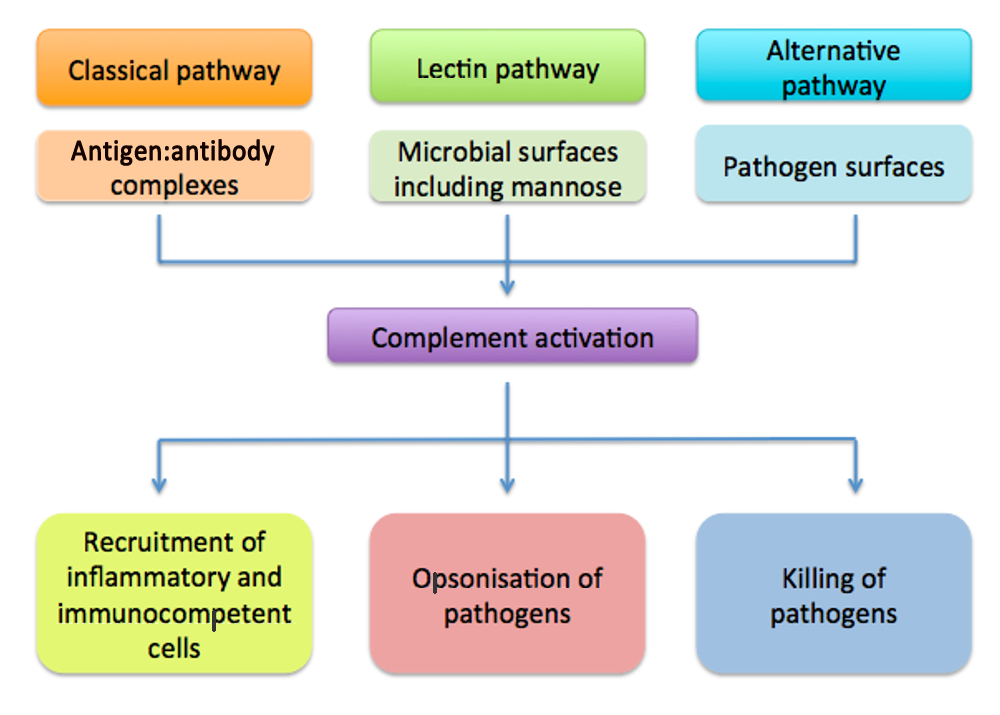
  • Complement pathway consists of three main pathways: classical, alternative and mannose binding lectin pathway.
  • All pathways converge on one common final or terminal pathway which generates membrane attack complex.

Infection associated/typical PID

There are a group of complement disorders that present with similar frequent infections as with other PID (Frank, 2010).

However, HAE does not present with the classic PID frequency of infection.  
Deficiencies in classical pathway

  • Classical pathway important in clearing of immune complexes and dead/dying cells (Truedsson, 2015).
  • Primary deficiency of C1, C2 or C4 is linked to development of SLE.
  • C2 deficiency most common complement deficiency in 1:20 000 (Sjöholm et al., 2006).
  • Hereditary angioedema (HAE) due to C1 inhibitor deficiency, a control protein of classical pathway.

Deficiencies in alternative pathway

  • Factor B and D deficiency are exceedingly rare (Kavanagh et al., 2025).
  • Properidin deficiency is X linked and associated with increased susceptibility to Niesserial infections (Agarwal et al., 2010).
  • Deficiencies of alternative pathway control proteins may cause consumption of complement and increase risks of infection and autoimmunity. 
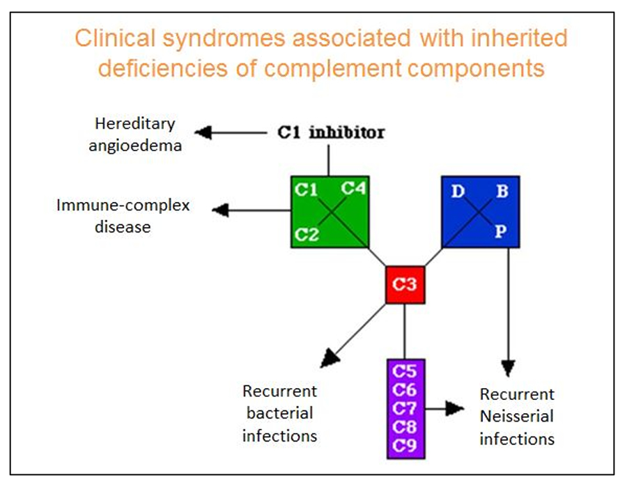
Clinical syndromes 

Deficiencies in mannose lectin pathway

  • MBL binds to carbohydrates on microbes or unwanted material and triggers the activation of the lectin pathway (Takahashi et al., 2006).
  • No consensus on the clinical relevance of MBL deficiency.
  • The majority of individuals with low MBL levels are asymptomatic.
  • However, increased susceptibility to infection with MBL deficiency appears to occur when there are other concomitant factors impairing the immune system.

Deficiencies in common final pathway

  • Major feature in this group is recurrent infections (Xie et al., 2020).
  • Generates membrane attack complex.
  • Infection with Neisseria meningitides particularly common. Rare serotypes such a W135 and Y may cause disease. 

Investigation of complement deficiency

  • Clinical indications for investigation include recurrent bacterial infections and autoimmunity (Coss et al., 2023).
  • HAE patients do not present with recurrent infections.
  • Investigation of complement deficiency in consultation with clinical immunologist/specialist (McMurray et al., 2024):
    • Initial screen for complement is C3, C4 levels and CH50 and may include AH50.
    • CH50 assesses classical pathway and final common pathway.
    • AH50 assesses alternative and final common pathway.
    • Low CH50 alone suggests deficiency of C1, C2 or C4.
    • Low AH50 alone suggests deficiency of Factor B, Factor D or Properidin.
    • Low AH50 and CH50 suggests deficiency of C3 or final common pathway.

Hereditary Angioedema (HAE)

  • Rare autosomal dominant genetic disorder due to deficiency of C1 inhibitor (C1 INH) protein (Campos et al., 2021).
  • Patients suffer unpredictable and recurrent episodes of angioedema involving skin, gut and airway.
  • Angioedema results from excessive production of bradykinin, a potent vasodilatory mediator (Germenis et al., 2020).
  • Histamine and other mast cell mediators are not directly involved, which explains the lack of response to antihistamines.
  • An acquired form of C1 INH deficiency may be seen in older patients without a family history of angioedema and is associated with underlying disorders and autoantibodies in most cases. Acquired C1-INH deficiency is not discussed further in this module (Trainotti et al., 2023).

Types of HAE

Type 1

  • Type 1 HAE most common (85%) due to low levels of the C1 inhibitor (C1-INH) (Guan et al., 2024).
  • Upon testing, protein (antigenic) and functional levels are both low.

Type 2

  • Type 2 HAE (remaining 15%) due to normal levels but abnormal function of C1-INH (Abuzakouk et al., 2018).
  • Upon testing normal or elevated protein levels but functional levels are low.

Type 3

  • Distinguished from other types of HAE due to normal levels and function of C1 inhibitor (Miranda et al., 2013).
  • Some cases associated with mutations of Factor XII

In both Type 1 and Type 2 cases, C4 levels are usually low due to consumption of complement. 

Indications for screening

Indications for screening of C1 inhibitor disorders include the presence of one or more of the following (Gülbahar, 2021):

  • Recurrent angioedema without urticaria.
  • Unexplained recurrent episodes of self-limited, colicky, abdominal pain.
  • A family history of angioedema.
  • Unexplained laryngeal oedema (even a single episode).
  • Low C4 levels, especially in the setting of angioedema.

Diagnosis

  • C1 Inhibitor protein (antigenic) and functional levels should be assessed (Farkas et al., 2017).
  • Abnormal results should be repeated to confirm.
  • Low C4 level would be supportive.
  • All diagnosed patients should be referred to a clinical immunologist with experience in managing HAE patients.
  • Genetic testing is not required to confirm the diagnosis in adults.
  • C1-INH levels are difficult to interpret in children younger than one year.

Testing family members

  • Once a diagnosis of HAE has been made, testing of the patient’s children, parents, and siblings (if parents carry the mutation) should be encouraged (Fragnan et al., 2018).
  • Although autosomal dominant approximately 25% of cases result from de novo mutations, so may not always have affected family members.
  • Affected individuals may be asymptomatic, especially early in childhood.

HAE attacks

  • Attack frequency is variable (Mendivil et al., 2023).
  • Untreated patients may have attacks up to every one to two weeks.
  • Swelling usually builds up over 24 hours, and may last three to four days or longer.
  • Manifestations:
  • Abdominal pain – nausea, vomiting, dehydration, diarrhoea or constipation.
  • Swelling – face, limbs, genitals.
  • Oropharangeal swelling – less frequent, but life threatening.
  • No itching or urticaria.
  • Does not respond to adrenaline, antihistamine or corticosteroids. 

Prodromal symptoms

Occurs in up to 50% of HAE patients (Prematta et al., 2009; Magerl et al., 2018):

  • Fatigue.
  • Flu-like symptoms.
  • Indigestion.
  • Tingling or “tightness” sensation at site where swelling then occurs.
  • About 25% of patients will have a flat, non-itching, blotchy, red rash before and during the attack.

Triggers of HAE include:

  • Anxiety and stress
  • Minor trauma or injury
  • Some surgical and dental procedures
  • Oestrogens
  • ACE inhibitors
  • Infections
  • Sometimes no trigger factor can be identified

Zarnowski and Treudler (2024)

Treatment options for HAE

Drug

Action

Route

Acute treatment

Berinert

Cinryze*

C1-inhibitor concentrate

IV

Firazyr (icatibant)**

B2 bradykinin receptor antagonist

SC

Prophylaxis

Danazol

Anabolic steroid

Oral

Cinryze*

C1-inhibitor concentrate

IV

Tranexamic Acid

Antifibrinolytic

Oral

*Not registered in NZ

** Not licensed for children below 12 years

Valerieva and Longhurst (2022)

Ongoing care

  • Education about HAE is very important, particularly in newly diagnosed patients (Riedl et al., 2022).
  • Trigger avoidance – triggers vary, but generally avoidance of trauma particularly to the face and upper respiratory tract and recognition and prompt treatment of oral and dental infections.
  • Planning for acute treatment – all patients should be equipped with:
  • Vaccinations – patients should be vaccinated against hepatitis B in case they require blood-derived products in the future (Craig et al., 2012).

Indications for prophylaxis

Short term prophylaxis should always be administered if intubation, oral surgery or general surgery is planned, as oral surgery is one of the most predictable triggers for HAE attacks (Farkas et al., 2012).

Long term prophylaxis is given to decrease the overall number of attacks (Li, 2020). 

Further information

ASCIA HAE position statement and action plan

www.allergy.org.au/health-professionals/papers/hereditary-angioedema

HAE Australasia www.haeaustralasia.org.au/ 

7. Other PID syndromes

DiGeorge Syndrome

DiGeorge syndrome:

  • Is due to defective embryonic development of the 3rd and 4th pharyngeal pouches, with the classic features of conotruncal abnormalities, thymic hypoplasia and hypoparathyroidism (Fomin et al., 2010).
  • Is often associated with 22q11.2 deletion (which affects 1:4000 live births) but absence of 22q22.1 deletion does not exclude DiGeorge syndrome (De decker and Lawrenson, 2001) .
  • May present with a wide phenotypic spectrum.

The spectrum of immune defect in patients with DiGeorge syndrome varies widely and is not related to other clinical features (McLean-Took et al., 2007):

  • Most commonly mild to moderate decrease in T cells, and patients are not clinically immunodeficient.
  • Antibody deficiencies can sometimes be seen, and may necessitate antibiotic prophylaxis or immunoglobulin replacement therapy.
  • Autoimmunity can also be seen.
  • Complete DiGeorge syndrome affects <1% of patients, who have complete absence of thymus.  These patients have no T cell function and present with SCID phenotype, requiring HSCT or thymic transplant.
  • Hypoplasia of parathyroid glands is associated with hypocalcaemia.
  • Contruncal cardiac defects may occur including Tetralogy of Fallot, Ventricular septal defect and interrupted aortic arch.
  • Patients almost always show abnormal facial features but may be subtle.
  • 50% of patients will have ‘non typical’ malformations including opthalimic, renal, endocrine and musculoskeletal anomalies.

Diagnosis of DiGeorge syndrome involves karotyping and fluorescent in-situ hybridization (FISH) to look for 22q11.2 deletion (Larson and Butler, 1995).

Treatment of DiGeorge syndrome is dependent upon specific clinical features (Mustillo et al., 2023):

  • Hypocalcaemia may require calcium supplements.
  • Surgical correction of congenital heart defect usually needed.
  • Immune reconstitution is not recommended for milder immunological defects.
  • IVIg and antibiotic prophylaxis may be indicated dependent on immune function.

Wiskott-Aldrich Syndrome (WAS)

Wiskott-Aldrich (WAS) is an X-linked recessive disease due to mutation in the Wiskott-Aldrich Syndrome Protein (WASP) gene, which is important in cytoskeletal organisation of cells. Mutations can result in a wide spectrum of disease (Jin et al., 2004):

  • Classic WAS was originally described as a triad of immunodeficiency, thrombocytopenia with small platelets and eczema.  
  • Severe disease results in a broad immunodeficiency (affecting both T and B cells, which may worsen with time), autoimmunity and malignancy.
  • These patients may present with early opportunistic infection (e.g. PJP).
  • Milder forms include isolated thrombocytopenia (XLT) or neutropenia (XLN).

Investigations typically show thrombocytopenia with small platelets, low serum IgM and poor pneumococcal antibody responses (Sudhakar et al., 2021).

Treatment may include (Buchbinder et al., 2014):

  • Immunoglobulin replacement therapy and antibiotic prophylaxis may be indicated dependent on immune function.
  • Bleeding problems may require platelet transfusions.
  • Patients with severe disease require HSCT.

Transient hypogammaglobulinaemia of infancy (THI)

Development of immunoglobulins through infancy and early childhood follows a predictable pattern, with IgM reaching adult normal ranges in the first year of life, IgG by middle childhood, and IgA by adolescence (Justiz-Vaillant et al., 2023). However, in some infants and young children the production of immunoglobulins is delayed. These children may present with recurrent mild infections particularly in the first one to two years of life. The diagnosis of transient hypogammaglobulinaemia of infancy (THI) can only be securely made once immunoglobulin levels normalise, and these children need exclusion of more significant immunodeficiency.

Management for most children with THI involves periodic monitoring (e.g. six monthly) of immunoglobulin levels (Ameratunga et al., 2019). Prophylactic antibiotics may be needed, and rarely a defined period of immunoglobulin replacement may be considered, with a plan to reevaluate with time.

Chronic Granulomatous Disease (CGD)

Chronic Granulomatous Disease (CGD) is a rare disorder where neutrophils are able to ingest organisms but cannot kill them, resulting in granuloma formation (Roos, 2016). CGD is due to mutations in nicotinamide adenine dinucleotide phosphate (NADPH) oxidase complex which normally generates microbicidal respiratory bursts. CGD can present at any age, but most commonly during early childhood.

Patients with CGD are susceptible to recurrent and persistent infections with organisms which are resistant to non-oxidative killing. An X-linked mutation in gp91 component accounts for 65% of cases. Other causes are autosomal recessive, therefore girls can also be affected (Holland, 2013).

Patients with CGD usually present with (Staudacher and von Bernuth, 2024):

  • Infections - frequent first manifestations including pneumonia, cellulitis and skin abscesses, liver abscesses and osteomyelitis (usually caused by Staphylococcus aureus).
  • Fungal infections with Aspergillus may be seen as well as infections with unusual organisms such as mycobacteria and burkholderia.
  • Development of granulomas in the skin, gastrointestinal (GI) tract, and genitourinary (GU) tract.
  • Granulomatous inflammation of bowel which may cause granulomatous colitis, Crohns-like inflammatory bowel disease (IBD) or bowel obstruction.

Investigations include nitroblue tetrazolium (NBT) and/or Dihydrorhodamine (DHR) tests for neutrophil oxidase activity (Yu et al., 2018). Specialised protein or genetic tests are useful to establish subtype and genetic inheritance pattern.

Treatment options for CGD include (Oikonomopoulou et al., 2022):

  • Antibacterial and antifungal prophylaxis.
  • Gamma Interferon.
  • Haematopoetic stem cell transplant (HSCT).

Hemophagocytic lymphohistiocytosis (HLH)

Hemophagocytic lymphohistiocytosis (HLH) is a life threatening syndrome of excessive immune activation (Kaçar and Celkan, 2022).

HLH (George, 2014):

  • May occur as a primary (familial/hereditary) or secondary (non-familial/hereditary) disorder.
  • Is characterised by hereditary or acquired impaired cytotoxic function of NK cells and cytotoxic T-cells.
  • Impaired killing of target cells results in excess pro-inflammatory cytokine release (IFN-g, TNF-a, GM-CSF) from activated T cells and macrophages (cytokine storm).
  • Presentation of primary HLH is often triggered by infections (especially EBV or CMV) or other metabolic/immune stress.
  • Common genetic causes of primary HLH - Familial HLH (FHL) types 2-5 (perforin, MUNC13-4, syntaxin 11, syntaxin binding protein), X-linked lymphoproliferative syndrome (SAP (XLP1) or XIAP (XLP2)), Chediak-Higashi syndrome, Griscelli syndrome type 2, Hermansky-Pudlak syndrome type 2).
  • Primary HLH usually develops during the first one to three years of life but late-onset cases can present at any age.
  • Secondary HLH develops in association with infection, autoimmune / rheumatological conditions and malignancy.
  • Molecular mechanisms involved in secondary HLH are unknown but may be predisposed to by polymorphisms in or carrier status of genes associated with primary HLH.
  • Early diagnosis is important.
  • Typical presentation with persistent fever, hepatic and/or renal dysfunction, pancytopenia, splenomegaly, hemorrhagic diathesis, neurological symptoms. 

Diagnostic criteria for HLH

A. Molecular diagnosis consistent with HLH: pathologic mutations of PRF1, UNC13D, Munc18-2, Rab27a, STX11, SH2D1a, or BIRC4 (Chinnici et al., 2023)

or 

B. Five of the eight criteria below are fulfilled (Henter et al., 2024):

  1. Fever ≥ 38.5°C 
  2. Splenomegaly
  3. Cytopenias (affecting at least 2 of 3 lineages in the peripheral blood): 
    Hemoglobin <9 g/dL (in infants <4 weeks: hemoglobin <10 g/dL) 
    Platelets <100 x 103/ml           Neutrophils <1 103/ml 
  4. Hypertriglyceridemia (fasting, >265 mg/dL) and/or hypofibrinogenemia (<150 mg/dL)
  5. Hemophagocytosis in bone marrow, spleen, lymph nodes or liver
  6. Low or absent NK-cell activity
  7. Ferritin >500 ng/mL
  8. Elevated  sCD25 (α-chain of sIL-2 receptor)

HLH treatment

  • Early aggressive supportive therapy and treatment of infection essential.
  • Immunosuppressive therapy to control the hypercytokinemia with steroids, cyclosporin, intravenous immunoglobulin +/- etoposide or anti-thymocyte globulin (especially if primary HLH) - commonly used protocols are HLH-94 and HLH-2004 (Wu et al., 2024).
  • Allogeneic hematopoietic stem-cell transplantation (HSCT) recommended for primary HLH or refractory secondary disease (Lehmberg et al., 2019).
  • High morbidity and mortality (Marsh et al., 2010):
    • Death during acute phase in 10%-15%,
    • <10% 3 year survival if untreated,
    • 21-26% 5 year survival with treatment,
    • 43%-92% 3 year survival post HSCT. 

© ASCIA 2025

ASCIA is the peak professional body of clinical immunology/allergy specialists in Australia and New Zealand.

ASCIA resources are based on published literature and expert review, however, they are not intended to replace medical advice. The content of ASCIA resources is not influenced by any commercial organisations.

For more information go to www.allergy.org.au

To donate to immunology/allergy research go to www.allergyimmunology.org.au

8. References

For Reference please download the PDF pdfASCIA HP Clinical Update PID 2025669.69 KB.

Content updated May 2025

Primary immunodeficiencies (PID) Clinical Update old copy

ASCIA PID Clinical UpdateThis Clinical Update complements ASCIA primary immunodeficiencies (PID) e-training for health professionals.

The main purpose of this document and the ASCIA PID e-training course is to:

  • Increase awareness of PID assist health professionals, particularly general practitioners, paediatricians and general physicians.
  • Promote early recognition and referral to a clinical immunologist to improve the management and quality of life for patients with PID in Australia and New Zealand.


Contents include:

1. Immunodeficiency overview

2. Recognition, diagnosis and management of PID

3. Immunoglobulin Replacement Therapy (IRT)

4. Severe Combined Immunodeficiency (SCID)

5. Antibody deficiency disorders

6. Complement disorders including HAE

7. Other PID syndromes

8. References and further information

For further information go to https://traininghp.ascia.org.au/ to complete the ASCIA primary immunodeficiencies (PID) e-training for health professionals, which includes a final assessment.

© ASCIA 2025

Disclaimer: ASCIA information is reviewed by ASCIA members and represents the available published literature at the time of review. The content of this document is not intended to replace professional medical advice and any questions regarding a medical diagnosis or treatment should be directed to a medical practitioner. 

Content updated May 2025

Immunodeficiency References

pdfASCIA HP Immunodeficiency References 2025299.29 KB  

Abolhassani, H., Sadaghiani, M. S., Aghamohammadi, A., Ochs, H. D., & Rezaei, N. (2012). Home-based subcutaneous immunoglobulin versus hospital-based intravenous immunoglobulin in treatment of primary antibody deficiencies: systematic review and meta analysis. Journal of clinical immunology, 32, 1180-1192. https://doi.org/10.1007/s10875-012-9720-1  

Adrian YS Lee, P. E. G. (2014). Evaluating for immunodeficiency in children with recurrent infection. Australian Journal for General Practitioners, 43, 629-632. https://www.racgp.org.au/afp/2014/september/evaluating-for-immunodeficiency-in-children-with-r  

Agarwal, S., Ferreira, V. P., Cortes, C., Pangburn, M. K., Rice, P. A., & Ram, S. (2010). An evaluation of the role of properdin in alternative pathway activation on Neisseria meningitidis and Neisseria gonorrhoeae. J Immunol, 185(1), 507-516. https://doi.org/10.4049/jimmunol.0903598  

Åhlin, A., Fugeläng, J., de Boer, M., Ringden, O., Fasth, A., & Winiarski, J. (2013). Chronic granulomatous disease-haematopoietic stem cell transplantation versus conventional treatment. Acta Paediatr, 102(11), 1087-1094. https://doi.org/10.1111/apa.12384  

Akalu, Y. T., & Bogunovic, D. (2024). Inborn errors of immunity: an expanding universe of disease and genetic architecture. Nature Reviews Genetics, 25(3), 184-195. https://doi.org/10.1038/s41576-023-00656-z  

Al-Herz, W., Bousfiha, A., Casanova, J.-L., Chatila, T., Conley, M. E., Cunningham-Rundles, C., Etzioni, A., Franco, J. L., Gaspar, H. B., Holland, S. M., Klein, C., Nonoyama, S., Ochs, H. D., Oksenhendler, E., Picard, C., Puck, J. M., Sullivan, K., & Tang, M. L. K. (2014). Primary Immunodeficiency Diseases: An Update on the Classification from the International Union of Immunological Societies Expert Committee for Primary Immunodeficiency [Classification]. Frontiers in Immunology, Volume 5 - 2014. https://doi.org/10.3389/fimmu.2014.00162  

Ameratunga, R., Ahn, Y., Steele, R., & Woon, S. T. (2019). Transient hypogammaglobulinaemia of infancy: many patients recover in adolescence and adulthood. Clinical and Experimental Immunology, 198(2), 224-232. https://doi.org/10.1111/cei.13345  

Ameratunga, R., Woon, S.-T., Brewerton, M., Koopmans, W., Jordan, A., Brothers, S., & Singh, R. (2011). Primary immune deficiency disorders in the South Pacific: the clinical utility of a customized genetic testing program in New Zealand. Annals of the New York Academy of Sciences, 1238(1), 53-64. https://doi.org/10.1111/j.1749-6632.2011.06238.x   

Ameratunga, R., Woon, S.-T., Gillis, D., Koopmans, W., & Steele, R. (2013). New diagnostic criteria for common variable immune deficiency (CVID), which may assist with decisions to treat with intravenous or subcutaneous immunoglobulin. Clinical and Experimental Immunology, 174(2), 203-211. https://doi.org/10.1111/cei.12178  

Anderson, J. T., Cowan, J., Condino-Neto, A., Levy, D., & Prusty, S. (2022). Health-related quality of life in primary immunodeficiencies: Impact of delayed diagnosis and treatment burden. Clinical Immunology, 236, 108931. https://doi.org/10.1016/j.clim.2022.108931  

APIIEG. (2009). Recommendations for the use of Immunoglobulin Replacement Therapy in Immune Deficiency.  . http://www.korektorzdrowia.pl/wp-content/uploads/apiieg_2009.pdf  

Aranda, C. S., Gouveia-Pereira, M. P., da Silva, C. J. M., Rizzo, M. C. F. V., Ishizuka, E., de Oliveira, E. B., & Condino-Neto, A. (2024). Severe combined immunodeficiency diagnosis and genetic defects. Immunological Reviews, 322(1), 138-147. https://doi.org/10.1111/imr.13310   

Arkwright, P. D., & Gennery, A. R. (2011). Ten warning signs of primary immunodeficiency: a new paradigm is needed for the 21st century. Annals of the New York Academy of Sciences, 1238(1), 7-14. https://doi.org/10.1111/j.1749-6632.2011.06206.x  

ASCIA-TAPID. (2019). Consensus Guideline: Diagnosis, management and transplantation of severe combined immunodeficiency in Australia and New Zealand. . https://www.allergy.org.au/hp/papers/ascia-guidelines-scid-tapid

Australian Technical Advisory Group on Immunisation (ATAGI). Australian Immunisation Handbook, Australian Government Department of Health and Aged Care, Canberra, 2022, https://immunisationhandbook.health.gov.au  

Ballow, M., Sánchez-Ramón, S., & Walter, J. E. (2022). Secondary Immune Deficiency and Primary Immune Deficiency Crossovers: Hematological Malignancies and Autoimmune Diseases. Front Immunol, 13, 928062. https://doi.org/10.3389/fimmu.2022.928062   

Berger, M. (2011). Incidence of Infection is Inversely Related to Steady-State (Trough) Serum IgG Level in Studies of Subcutaneous IgG in PIDD. Journal of clinical immunology, 31(5), 924-926. https://doi.org/10.1007/s10875-011-9546-2  

Bezrodnik, L., Gómez Raccio, A., Belardinelli, G., Regairaz, L., Díaz Ballve, D., Seminario, G., Moreira, I., Riganti, C., Cantisano, C., Díaz, H., & Di Giovanni, D. (2013). Comparative Study of Subcutaneous Versus Intravenous IgG Replacement Therapy in Pediatric Patients with Primary Immunodeficiency Diseases: A Multicenter Study in Argentina. Journal of clinical immunology, 33(7), 1216-1222. https://doi.org/10.1007/s10875-013-9916-z   

Bonilla, F. A., Barlan, I., Chapel, H., Costa-Carvalho, B. T., Cunningham-Rundles, C., de la Morena, M. T., Espinosa-Rosales, F. J., Hammarström, L., Nonoyama, S., Quinti, I., Routes, J. M., Tang, M. L. K., & Warnatz, K. (2016). International Consensus Document (ICON): Common Variable Immunodeficiency Disorders. The Journal of Allergy and Clinical Immunology: In Practice, 4(1), 38-59. https://doi.org/10.1016/j.jaip.2015.07.025  

Bonilla, F. A., Khan, D. A., Ballas, Z. K., Chinen, J., Frank, M. M., Hsu, J. T., Keller, M., Kobrynski, L. J., Komarow, H. D., Mazer, B., Nelson, R. P., Jr., Orange, J. S., Routes, J. M., Shearer, W. T., Sorensen, R. U., Verbsky, J. W., Bernstein, D. I., Blessing-Moore, J., Lang, D.,…Verbsky, J. W. (2015). Practice parameter for the diagnosis and management of primary immunodeficiency. Journal of Allergy and Clinical Immunology, 136(5), 1186-1205.e1178. https://doi.org/10.1016/j.jaci.2015.04.049  

Bousfiha, A., Moundir, A., Tangye, S. G., Picard, C., Jeddane, L., Al-Herz, W., Rundles, C. C., Franco, J. L., Holland, S. M., Klein, C., Morio, T., Oksenhendler, E., Puel, A., Puck, J., Seppänen, M. R. J., Somech, R., Su, H. C., Sullivan, K. E., Torgerson, T. R., & Meyts, I. (2022). The 2022 Update of IUIS Phenotypical Classification for Human Inborn Errors of Immunity. Journal of clinical immunology, 42(7), 1508-1520. https://doi.org/10.1007/s10875-022-01352-z  

Bousfiha, A. A., Jeddane, L., Moundir, A., Poli, M. C., Aksentijevich, I., Cunningham-Rundles, C., Hambleton, S., Klein, C., Morio, T., Picard, C., Puel, A., Rezaei, N., Seppänen, M. R. J., Somech, R., Su, H. C., Sullivan, K. E., Torgerson, T. R., Tangye, S. G., & Meyts, I. (2025). The 2024 update of IUIS phenotypic classification of human inborn errors of immunity. Journal of Human Immunity, 1(1), e20250002. https://doi.org/10.70962/jhi.20250002  

Bucciol, G., Delafontaine, S., Meyts, I., & Poli, C. (2024). Inborn errors of immunity: A field without frontiers. Immunological Reviews, 322(1), 15-27. https://doi.org/10.1111/imr.13297  

Buchbinder, D., Nugent, D. J., & Fillipovich, A. H. (2014). Wiskott-Aldrich syndrome: diagnosis, current management, and emerging treatments. Appl Clin Genet, 7, 55-66. https://doi.org/10.2147/tacg.S58444  

Buckley, R. H. (2002). Primary cellular immunodeficiencies. J Allergy Clin Immunol, 109(5), 747-757. https://doi.org/10.1067/mai.2002.123617  

Cardenas-Morales, M., & Hernandez-Trujillo, V. P. (2022). Agammaglobulinemia: from X-linked to Autosomal Forms of Disease. Clin Rev Allergy Immunol, 63(1), 22-35. https://doi.org/10.1007/s12016-021-08870-5  

Casanova, J.-L., & Abel, L. (2007). Primary Immunodeficiencies: A Field in Its Infancy. Science, 317(5838), 617-619. https://doi.org/doi:10.1126/science.1142963  

Casanova, J.-L., & Abel, L. (2022). From rare disorders of immunity to common determinants of infection: Following the mechanistic thread. Cell, 185(17), 3086-3103. https://doi.org/10.1016/j.cell.2022.07.004  

Casanova, J.-L., & Anderson, M. S. (2023). Unlocking life-threatening COVID-19 through two types of inborn errors of type I IFNs. The Journal of Clinical Investigation, 133(3). https://doi.org/10.1172/JCI166283   

Casanova, J.-L., Conley, M. E., Seligman, S. J., Abel, L., & Notarangelo, L. D. (2014). Guidelines for genetic studies in single patients: lessons from primary immunodeficiencies. Journal of Experimental Medicine, 211(11), 2137-2149. https://doi.org/10.1084/jem.20140520  

Castagnoli, R., Delmonte, O. M., Calzoni, E., & Notarangelo, L. D. (2019). Hematopoietic Stem Cell Transplantation in Primary Immunodeficiency Diseases: Current Status and Future Perspectives. Front Pediatr, 7, 295. https://doi.org/10.3389/fped.2019.00295  

Cavazzana-Calvo, M., Hacein-Bey, S., Basile, G. d. S., Gross, F., Yvon, E., Nusbaum, P., Selz, F., Hue, C., Certain, S., Casanova, J.-L., Bousso, P., Deist, F. L., & Fischer, A. (2000). Gene therapy of human severe combined immunodeficiency (SCID)-X1 disease. Science, 288(5466), 669-672. https://doi.org/doi:10.1126/science.288.5466.669  

Chan, A., Scalchunes, C., Boyle, M., & Puck, J. M. (2011). Early vs. delayed diagnosis of severe combined immunodeficiency: a family perspective survey. Clin Immunol, 138(1), 3-8. https://doi.org/10.1016/j.clim.2010.09.010  

Chapel, H., Prevot, J., Gaspar, H. B., Español, T., Bonilla, F. A., Solis, L., & Drabwell, J. (2014). Primary Immune Deficiencies – Principles of Care [Hypothesis and Theory]. Frontiers in Immunology, Volume 5 - 2014. https://doi.org/10.3389/fimmu.2014.00627

Chen, R., Lukianova, E., van der Loeff, I. S., Spegarova, J. S., Willet, J. D. P., James, K. D., Ryder, E. J., Griffin, H., IJspeert, H., Gajbhiye, A., Lamoliatte, F., Marin-Rubio, J. L., Woodbine, L., Lemos, H., Swan, D. J., Pintar, V., Sayes, K., Ruiz-Morales, E. R., Eastham, S.,…Hambleton, S. (2024). NUDCD3 deficiency disrupts V(D)J recombination to cause SCID and Omenn syndrome. Science Immunology, 9(95), eade5705. https://doi.org/doi:10.1126/sciimmunol.ade5705  

Chinnici, A., Beneforti, L., Pegoraro, F., Trambusti, I., Tondo, A., Favre, C., Coniglio, M. L., & Sieni, E. (2023). Approaching hemophagocytic lymphohistiocytosis [Review]. Frontiers in Immunology, Volume 14 - 2023. https://doi.org/10.3389/fimmu.2023.1210041  

Chong-Neto, H. J., Radwan, N., Condino-Neto, A., Rosário Filho, N. A., Ortega-Martell, J. A., & El-Sayed, Z. A. (2024). Newborn screening for inborn errors of immunity: The status worldwide. World Allergy Organ J, 17(6), 100920. https://doi.org/10.1016/j.waojou.2024.100920  

Cinetto, F., Neri, R., Vianello, F., Visentin, A., Barilà, G., Gianese, S., Lanciarotta, A., Milito, C., Rattazzi, M., Piazza, F., Trentin, L., Zambello, R., Agostini, C., & Scarpa, R. (2021). Subcutaneous immunoglobulins replacement therapy in secondary antibody deficiencies: Real life evidence as compared to primary antibody deficiencies. PLoS One, 16(3), e0247717. https://doi.org/10.1371/journal.pone.0247717  

Cole, T., McLean‐Tooke, A., Loh, R., Quinn, P., Peake, J., Sinclair, J., & Smart, J. . (2018). P48: ASCIA TRANSPLANTATION AND PRIMARY IMMUNODEFICIENCY (TAPID) PROJECT. Internal Medicine Journal, 48(S6), 18-19. https://doi.org/10.1111/imj.48_14077  

Cole, T., Pearce, M. S., Cant, A. J., Cale, C. M., Goldblatt, D., & Gennery, A. R. (2013). Clinical outcome in children with chronic granulomatous disease managed conservatively or with hematopoietic stem cell transplantation. Journal of Allergy and Clinical Immunology, 132(5), 1150-1155. https://doi.org/10.1016/j.jaci.2013.05. ng the Lives of People With Primary Immunodeficiencies (PI) With Early Testing and Diagnosis. Front Immunol, 9, 1439. https://doi.org/10.3389/fimmu.2018.01439  

Cooper, M. A., Pommering, T. L., & Korányi, K. (2003). Primary immunodeficiencies. Am Fam Physician, 68(10), 2001-2008.

Coss, S. L., Zhou, D., Chua, G. T., Aziz, R. A., Hoffman, R. P., Wu, Y. L., Ardoin, S. P., Atkinson, J. P., & Yu, C.-Y. (2023). The complement system and human autoimmune diseases. Journal of Autoimmunity, 137, 102979. https://doi.org/10.1016/j.jaut.2022.102979  

Cunningham-Rundles, C. (2012). The many faces of common variable immunodeficiency. Hematology Am Soc Hematol Educ Program, 2012, 301-305. https://doi.org/10.1182/asheducation-2012.1.301  

Dahl, C., Petersen, I., Ilkjær, F. V., Westh, L., Katzenstein, T. L., Hansen, A.-B. E., Nielsen, T. L., Larsen, C. S., Johansen, I. S., & Rasmussen, L. D. (2023). Missed Opportunities to Diagnose Common Variable Immunodeficiency: a Population-Based Case–Control Study Identifying Indicator Diseases for Common Variable Immunodeficiency. Journal of clinical immunology, 43(8), 2104-2114. https://doi.org/10.1007/s10875-023-01590-9  

De Decker, H. P., & Lawrenson, J. B. (2001). The 22q11.2 deletion: From diversity to a single gene theory. Genetics in Medicine, 3(1), 2-5. https://doi.org/10.1097/00125817-200101000-00002  

Dvorak, C. C., Haddad, E., Heimall, J., Dunn, E., Buckley, R. H., Kohn, D. B., Cowan, M. J., Pai, S. Y., Griffith, L. M., Cuvelier, G. D. E., Eissa, H., Shah, A. J., O'Reilly, R. J., Pulsipher, M. A., Wright, N. A. M., Abraham, R. S., Satter, L. F., Notarangelo, L. D., & Puck, J. M. (2023). The diagnosis of severe combined immunodeficiency (SCID): The Primary Immune Deficiency Treatment Consortium (PIDTC) 2022 Definitions. J Allergy Clin Immunol, 151(2), 539-546. https://doi.org/10.1016/j.jaci.2022.10.022  

EBMT/ESID. (2017). EBMT/ESID Guidelines for haematopoietic stem cell transplantation (HSCT) for primary immunodeficiencies HSCT Guidelines. https://www.ebmt.org/ebmt/documents/esid-ebmt-hsct-guidelines-2017

Fernández-Cruz, E., Alecsandru, D., & Ramón, S. S. (2009). Mechanisms of action of immune globulin. Clinical and Experimental Immunology, 157(Supplement_1), 1-2. https://doi.org/10.1111/j.1365-2249.2009.03955.x  

Firatoglu, H., Aytekin, C., Dogu, F., Bal, S. K., Haskologlu, S., Boztug, K., & Ikinciogullari, A. (2025). Evaluation of Patients with Combined Immunodeficiency: A Single Center Experience. Iran J Immunol, 22(1), 89-99. https://doi.org/10.22034/iji.2025.103499.2844  

Fischer, A. (2000). Severe combined immunodeficiencies (SCID). Clin Exp Immunol, 122(2), 143-149. https://doi.org/10.1046/j.1365-2249.2000.01359.x  

Fischer, A., & Rausell, A. (2018). What do primary immunodeficiencies tell us about the essentiality/redundancy of immune responses? Seminars in Immunology, 36, 13-16. https://doi.org/10.1016/j.smim.2017.12.001   

Fomin, A. B., Pastorino, A. C., Kim, C. A., Pereira, C. A., Carneiro-Sampaio, M., & Abe-Jacob, C. M. (2010). DiGeorge Syndrome: a not so rare disease. Clinics (Sao Paulo), 65(9), 865-869. https://doi.org/10.1590/s1807-59322010000900009  

Fornes, O., Jia, A., Kuehn, H. S., Min, Q., Pannicke, U., Schleussner, N., Thouenon, R., Yu, Z., de los Angeles Astbury, M., Biggs, C. M., Galicchio, M., Garcia-Campos, J. A., Gismondi, S., Gonzalez Villarreal, G., Hildebrand, K. J., Hönig, M., Hou, J., Moshous, D., Pittaluga, S.,…Wang, J.-Y. (2023). A multimorphic mutation in IRF4 causes human autosomal dominant combined immunodeficiency. Science Immunology, 8(79), eade7953. https://doi.org/doi:10.1126/sciimmunol.ade7953  

Frank, M. M. (2010). Complement disorders and hereditary angioedema. J Allergy Clin Immunol, 125(2 Suppl 2), S262-271. https://doi.org/10.1016/j.jaci.2009.10.063  

Gardulf, A. (2007). Immunoglobulin treatment for primary antibody deficiencies: advantages of the subcutaneous route. BioDrugs, 21(2), 105-116. https://doi.org/10.2165/00063030-200721020-00005   

Gaspar, J., Gerritsen, B., & Jones, A. (1998). Immunoglobulin replacement treatment by rapid subcutaneous infusion. Archives of Disease in Childhood, 79(1), 48. https://doi.org/10.1136/adc.79.1.48  

George, M. R. (2014). Hemophagocytic lymphohistiocytosis: review of etiologies and management. J Blood Med, 5, 69-86. https://doi.org/10.2147/jbm.S46255  

Gray, P. E. A., Namasivayam, M., & Ziegler, J. B. (2012). Recurrent infection in children: When and how to investigate for primary immunodeficiency? Journal of Paediatrics and Child Health, 48(3), 202-209. https://doi.org/10.1111/j.1440-1754.2011.02080.x  

Grosserichter-Wagener, C., Franco-Gallego, A., Ahmadi, F., Moncada-Vélez, M., Dalm, V. A. S. H., Rojas, J. L., Orrego, J. C., Correa Vargas, N., Hammarström, L., Schreurs, M. W. J., Dik, W. A., van Hagen, P. M., Boon, L., van Dongen, J. J. M., van der Burg, M., Pan-Hammarström, Q., Franco, J. L., & van Zelm, M. C. (2020). Defective formation of IgA memory B cells, Th1 and Th17 cells in symptomatic patients with selective IgA deficiency. Clinical & Translational Immunology, 9(5), e1130. https://doi.org/10.1002/cti2.1130  

Gruber, C., & Bogunovic, D. (2020). Incomplete penetrance in primary immunodeficiency: a skeleton in the closet. Hum Genet, 139(6-7), 745-757. https://doi.org/10.1007/s00439-020-02131-9  

Guérin, A., Kerner, G., Marr, N., Markle, J. G., Fenollar, F., Wong, N., Boughorbel, S., Avery, D. T., Ma, C. S., Bougarn, S., Bouaziz, M., Béziat, V., Della Mina, E., Oleaga-Quintas, C., Lazarov, T., Worley, L., Nguyen, T., Patin, E., Deswarte, C.,…Casanova, J.-L. (2018). IRF4 haploinsufficiency in a family with Whipple’s disease. eLife, 7, e32340. https://doi.org/10.7554/eLife.32340  

Guo, Y., Tian, X., Wang, X., & Xiao, Z. (2018). Adverse Effects of Immunoglobulin Therapy. Front Immunol, 9, 1299. https://doi.org/10.3389/fimmu.2018.01299  

Heather, N., de Hora, M., Brothers, S., Grainger, P., Knoll, D., & Webster, D. (2022). Introducing Newborn Screening for Severe Combined Immunodeficiency-The New Zealand Experience. Int J Neonatal Screen, 8(2). https://doi.org/10.3390/ijns8020033   

Heimall, J., & Cowan, M. J. (2017). Long term outcomes of severe combined immunodeficiency: therapy implications. Expert Rev Clin Immunol, 13(11), 1029-1040. https://doi.org/10.1080/1744666x.2017.1381558  

Henter, J. I., Sieni, E., Eriksson, J., Bergsten, E., Hed Myrberg, I., Canna, S. W., Coniglio, M. L., Cron, R. Q., Kernan, K. F., Kumar, A. R., Lehmberg, K., Minoia, F., Naqvi, A., Ravelli, A., Tang, Y. M., Bottai, M., Bryceson, Y. T., Horne, A., & Jordan, M. B. (2024). Diagnostic guidelines for familial hemophagocytic lymphohistiocytosis revisited. Blood, 144(22), 2308-2318. https://doi.org/10.1182/blood.2024025077  

Holland, S. M. (2013). Chronic granulomatous disease. Hematol Oncol Clin North Am, 27(1), 89-99, viii. https://doi.org/10.1016/j.hoc.2012.11.002

Houghton, B. C., & Booth, C. (2021). Gene Therapy for Primary Immunodeficiency. Hemasphere, 5(1), e509. https://doi.org/10.1097/hs9.0000000000000509  

Inwald, D. P., Peters, M. J., Walshe, D., Jones, A., Davies, E. G., & Klein, N. J. (2000). Absence of platelet CD40L identifies patients with X-linked hyper IgM syndrome. Clin Exp Immunol, 120(3), 499-502. https://doi.org/10.1046/j.1365-2249.2000.01235.x  

Israel, L., Wang, Y., Bulek, K., Della Mina, E., Zhang, Z., Pedergnana, V., Chrabieh, M., Lemmens, N. A., Sancho-Shimizu, V., Descatoire, M., Lasseau, T., Israelsson, E., Lorenzo, L., Yun, L., Belkadi, A., Moran, A., Weisman, L. E., Vandenesch, F., Batteux, F.,…Puel, A. (2017). Human Adaptive Immunity Rescues an Inborn Error of Innate Immunity. Cell, 168(5), 789-800.e710. https://doi.org/10.1016/j.cell.2017.01.039  

Jin, Y., Mazza, C., Christie, J. R., Giliani, S., Fiorini, M., Mella, P., Gandellini, F., Stewart, D. M., Zhu, Q., Nelson, D. L., Notarangelo, L. D., & Ochs, H. D. (2004). Mutations of the Wiskott-Aldrich Syndrome Protein (WASP): hotspots, effect on transcription, and translation and phenotype/genotype correlation. Blood, 104(13), 4010-4019. https://doi.org/10.1182/blood-2003-05-1592  

Johnson, M. B., Ogishi, M., Domingo-Vila, C., De Franco, E., Wakeling, M. N., Imane, Z., Resnick, B., Williams, E., Galão, R. P., Caswell, R., Russ-Silsby, J., Seeleuthner, Y., Rinchai, D., Fagniez, I., Benson, B., Dufort, M. J., Speake, C., Smithmyer, M. E., Hudson, M.,…Oram, R. A. (2024). Human inherited PD-L1 deficiency is clinically and immunologically less severe than PD-1 deficiency. J Exp Med, 221(6). https://doi.org/10.1084/jem.20231704  

Jolles, S., Orange, J. S., Gardulf, A., Stein, M. R., Shapiro, R., Borte, M., & Berger, M. (2015). Current treatment options with immunoglobulin G for the individualization of care in patients with primary immunodeficiency disease. Clinical and Experimental Immunology, 179(2), 146-160. https://doi.org/10.1111/cei.12485  

Jolles, S., Sewell, W. A., & Misbah, S. A. (2005). Clinical uses of intravenous immunoglobulin. Clin Exp Immunol, 142(1), 1-11. https://doi.org/10.1111/j.1365-2249.2005.02834.x

Jolles, S., & Sleasman, J. W. (2011). Subcutaneous immunoglobulin replacement therapy with Hizentra®, the first 20% SCIG preparation: a Practical approach. Advances in Therapy, 28(7), 521-533. https://doi.org/10.1007/s12325-011-0036-y  

Justiz-Vaillant, A. A., Hoyte, T., Davis, N., Deonarinesingh, C., De Silva, A., Dhanpaul, D., Dookhoo, C., Doorpat, J., Dopson, A., Durgapersad, J., Palmer, C., Asin-Milan, O., Williams-Persad, A. F.-A., & Arozarena-Fundora, R. (2023). A Systematic Review of the Clinical Diagnosis of Transient Hypogammaglobulinemia of Infancy. Children, 10(8), 1358. https://www.mdpi.com/2227-9067/10/8/1358  

Kaçar, A. G., & Celkan, T. T. (2022). Hemophagocytic Lymphohistiocytosis. Balkan Med J, 39(5), 309-317. https://doi.org/10.4274/balkanmedj.galenos.2022.2022-4-83  

Kang, E. M., Marciano, B. E., DeRavin, S., Zarember, K. A., Holland, S. M., & Malech, H. L. (2011). Chronic granulomatous disease: Overview and hematopoietic stem cell transplantation. Journal of Allergy and Clinical Immunology, 127(6), 1319-1326. https://doi.org/10.1016/j.jaci.2011.03.028  

Karhan, A. N., Esenboğa, S., Gümüş, E., Karaatmaca, B., Cagdas, D., Demir, H., Saltik Temizel İ, N., Özen, H., Yüce, A., & Tezcan, İ. (2022). Nutritional status of children with primary immunodeficiency: A single center experience. Pediatr Int, 64(1), e14996. https://doi.org/10.1111/ped.14996  

Kavanagh, D., Barratt, J., Schubart, A., Webb, N. J. A., Meier, M., & Fakhouri, F. (2025). Factor B as a therapeutic target for the treatment of complement-mediated diseases. Front Immunol, 16, 1537974. https://doi.org/10.3389/fimmu.2025.1537974  

Kirkpatrick, P., & Riminton, S. (2007). Primary Immunodeficiency Diseases in Australia and New Zealand. Journal of clinical immunology, 27(5), 517-524. https://doi.org/10.1007/s10875-007-9105-z  

Kobrynski, L. (2012). Subcutaneous immunoglobulin therapy: a new option for patients with primary immunodeficiency diseases. Biologics, 6, 277-287. https://doi.org/10.2147/btt.S25188  

Kohn, D. B., Hershfield, M. S., Puck, J. M., Aiuti, A., Blincoe, A., Gaspar, H. B., Notarangelo, L. D., & Grunebaum, E. (2019). Consensus approach for the management of severe combined immune deficiency caused by adenosine deaminase deficiency. Journal of Allergy and Clinical Immunology, 143(3), 852-863. https://doi.org/10.1016/j.jaci.2018.08.024  

Kostel Bal, S., Giuliani, S., Block, J., Repiscak, P., Hafemeister, C., Shahin, T., Kasap, N., Ransmayr, B., Miao, Y., van de Wetering, C., Frohne, A., Jimenez Heredia, R., Schuster, M., Zoghi, S., Hertlein, V., Thian, M., Bykov, A., Babayeva, R., Bilgic Eltan, S.,…Boztug, K. (2023). Biallelic NFATC1 mutations cause an inborn error of immunity with impaired CD8+ T-cell function and perturbed glycolysis. Blood, 142(9), 827-845. https://doi.org/10.1182/blood.2022018303  

Kuo, C. Y., Garabedian, E., Puck, J., Cowan, M. J., Sullivan, K. E., Buckley, R. H., Cunningham-Rundles, C., Marsh, R., Candotti, F., & Kohn, D. B. (2020). Adenosine Deaminase (ADA)-Deficient Severe Combined Immune Deficiency (SCID) in the US Immunodeficiency Network (USIDNet) Registry. J Clin Immunol, 40(8), 1124-1131. https://doi.org/10.1007/s10875-020-00857-9  

Larson, R. S., & Butler, M. G. (1995). Use of fluorescence in situ hybridization (FISH) in the diagnosis of DiGeorge sequence and related diseases. Diagn Mol Pathol, 4(4), 274-278. https://doi.org/10.1097/00019606-199512000-00008  

Lee, T. K., Gereige, J. D., & Maglione, P. J. (2021). State-of-the-art diagnostic evaluation of common variable immunodeficiency. Ann Allergy Asthma Immunol, 127(1), 19-27. https://doi.org/10.1016/j.anai.2021.03.005  

Lehmberg, K., Moshous, D., & Booth, C. (2019). Haematopoietic Stem Cell Transplantation for Primary Haemophagocytic Lymphohistiocytosis. Front Pediatr, 7, 435. https://doi.org/10.3389/fped.2019.00435  

Leiding, J. W., & Forbes, L. R. (2019). Mechanism-Based Precision Therapy for the Treatment of Primary Immunodeficiency and Primary Immunodysregulatory Diseases. The Journal of Allergy and Clinical Immunology: In Practice, 7(3), 761-773. https://doi.org/10.1016/j.jaip.2018.12.017  

Li, Y., Yu, Z., Schenk, M., Lagovsky, I., Illig, D., Walz, C., Rohlfs, M., Conca, R., Muise, A. M., Snapper, S. B., Uhlig, H. H., Garty, B. Z., Klein, C., & Kotlarz, D. (2023). Human MD2 deficiency-an inborn error of immunity with pleiotropic features. J Allergy Clin Immunol, 151(3), 791-796.e797. https://doi.org/10.1016/j.jaci.2022.09.033  

Li, Z., & Mahmood, I. (2024). Immunoglobulin therapies for primary immunodeficiency diseases (part 2): considerations for dosing strategies. Immunotherapy, 16(13), 895-905. https://doi.org/10.1080/1750743x.2024.2382074   

Lingman-Framme, J., & Fasth, A. (2013). Subcutaneous Immunoglobulin for Primary and Secondary Immunodeficiencies: an Evidence-Based Review. Drugs, 73(12), 1307-1319. https://doi.org/10.1007/s40265-013-0094-3  

Loh, R. K., Vale, S., & McLean-Tooke, A. (2013). Quantitative serum immunoglobulin tests. Aust Fam Physician, 42(4), 195-198.

Lucas, C. L. (2024). Human genetic errors of immunity illuminate an adaptive arsenal model of rapid defenses. Trends in Immunology, 45(2), 113-126. https://doi.org/10.1016/j.it.2023.12.006  

Ma, C. S., & Tangye, S. G. (2019). Flow Cytometric-Based Analysis of Defects in Lymphocyte Differentiation and Function Due to Inborn Errors of Immunity [Review]. Frontiers in Immunology, Volume 10 - 2019. https://doi.org/10.3389/fimmu.2019.02108  

Mallick, R., Hahn, N., & Scalchunes, C. (2025). Immunoglobulin replacement therapy in patients with primary and secondary immunodeficiencies: impact of infusion method on immunoglobulin-specific perceptions of quality of life and treatment satisfaction. Allergy, Asthma & Clinical Immunology, 21(1), 2. https://doi.org/10.1186/s13223-024-00939-y  

Marsh, R. A., Vaughn, G., Kim, M.-O., Li, D., Jodele, S., Joshi, S., Mehta, P. A., Davies, S. M., Jordan, M. B., Bleesing, J. J., & Filipovich, A. H. (2010). Reduced-intensity conditioning significantly improves survival of patients with hemophagocytic lymphohistiocytosis undergoing allogeneic hematopoietic cell transplantation. Blood, 116(26), 5824-5831. https://doi.org/10.1182/blood-2010-04-282392  

Marshall, J. S., Warrington, R., Watson, W., & Kim, H. L. (2018). An introduction to immunology and immunopathology. Allergy, Asthma & Clinical Immunology, 14(2), 49. https://doi.org/10.1186/s13223-018-0278-1  

McCusker, C., Upton, J., & Warrington, R. (2018). Primary immunodeficiency. Allergy, Asthma & Clinical Immunology, 14(2), 61. https://doi.org/10.1186/s13223-018-0290-5  

McCusker, C., & Warrington, R. (2011). Primary immunodeficiency. Allergy, Asthma & Clinical Immunology, 7(1), S11. https://doi.org/10.1186/1710-1492-7-S1-S11  

McLean-Tooke, A., Spickett, G. P., & Gennery, A. R. (2007). Immunodeficiency and Autoimmunity in 22q11.2 Deletion Syndrome. Scandinavian Journal of Immunology, 66(1), 1-7. https://doi.org/https://doi.org/10.1111/j.1365-3083.2007.01949.x  

McMurray, J. C., Schornack, B. J., Weskamp, A. L., Park, K. J., Pollock, J. D., Day, W. G., Brockshus, A. T., Beakes, D. E., Schwartz, D. J., Mikita, C. P., & Pittman, L. M. (2024). Immunodeficiency: Complement disorders. Allergy Asthma Proc, 45(5), 305-309. https://doi.org/10.2500/aap.2024.45.240050  

Mickey, D., Camacho, J. V., Khan, A., & Kaufman, D. (2024). Immunodeficiency: Quantitative and qualitative phagocytic cell defects. Allergy Asthma Proc, 45(5), 299-304. https://doi.org/10.2500/aap.2024.45.240049  

Mitchell, R., Nivison-Smith, I., Anazodo, A., Tiedemann, K., Shaw, P., Teague, L., Fraser, C., Carter, T., Tapp, H., Alvaro, F., & O’Brien, T. A. (2013). Outcomes of hematopoietic stem cell transplantation in primary immunodeficiency: a report from the Australian and New Zealand Children's Haematology Oncology Group and the Australasian Bone Marrow Transplant Recipient Registry. Biology of Blood and Marrow Transplantation, 19(3), 338-343. https://doi.org/10.1016/j.bbmt.2012.11.619  

Mizoguchi, Y., & Okada, S. (2021). Inborn errors of STAT1 immunity. Current Opinion in Immunology, 72, 59-64. https://doi.org/10.1016/j.coi.2021.02.009  

Mustillo, P. J., Sullivan, K. E., Chinn, I. K., Notarangelo, L. D., Haddad, E., Davies, E. G., de la Morena, M. T., Hartog, N., Yu, J. E., Hernandez-Trujillo, V. P., Ip, W., Franco, J., Gambineri, E., Hickey, S. E., Varga, E., & Markert, M. L. (2023). Clinical Practice Guidelines for the Immunological Management of Chromosome 22q11.2 Deletion Syndrome and Other Defects in Thymic Development. J Clin Immunol, 43(2), 247-270. https://doi.org/10.1007/s10875-022-01418-y  

Neven, B., & Ferrua, F. (2020). Hematopoietic Stem Cell Transplantation for Combined Immunodeficiencies, on Behalf of IEWP-EBMT [Mini Review]. Frontiers in Pediatrics, Volume 7 - 2019. https://doi.org/10.3389/fped.2019.00552  

Nicolay, U., Haag, S., Eichmann, F., Herget, S., Spruck, D., & Gardulf, A. (2005). Measuring treatment satisfaction in patients with primary immunodeficiency diseases receiving lifelong immunoglobulin replacement therapy. Quality of Life Research, 14(7), 1683-1691. https://doi.org/10.1007/s11136-005-1746-x  

Notarangelo, L. D., Bacchetta, R., Casanova, J.-L., & Su, H. C. (2020). Human inborn errors of immunity: An expanding universe. Science Immunology, 5(49), eabb1662. https://doi.org/10.1126/sciimmunol.abb1662  

Ochs, H. D., Smith, C. E., & Puck, J. M. (2013). Primary Immunodeficiency Diseases: A Molecular and Genetic Approach. Oxford University Press. https://doi.org/10.1093/med/9780195389838.001.0001   

Ochs, H. D., Gupta, S., Kiessling, P., Nicolay, U., & Berger, M. (2006). Safety and Efficacy of Self-Administered Subcutaneous Immunoglobulin in Patients with Primary Immunodeficiency Diseases. Journal of clinical immunology, 26(3), 265-273. https://doi.org/10.1007/s10875-006-9021-7   

Oikonomopoulou, Z., Shulman, S., Mets, M., & Katz, B. (2022). Chronic Granulomatous Disease: an Updated Experience, with Emphasis on Newly Recognized Features. J Clin Immunol, 42(7), 1411-1419. https://doi.org/10.1007/s10875-022-01294-6  

O'Keefe, A. W., Halbrich, M., Ben-Shoshan, M., & McCusker, C. (2016). Primary immunodeficiency for the primary care provider. Paediatrics & Child Health, 21(2), e10-e14. https://doi.org/10.1093/pch/21.2.e10  

Oliveira, J. B., & Fleisher, T. A. (2010). Laboratory evaluation of primary immunodeficiencies. J Allergy Clin Immunol, 125(2 Suppl 2), S297-305. https://doi.org/10.1016/j.jaci.2009.08.043  

Ozdemir, O. (2021). Primary immunodeficiency diseases in the newborn. North Clin Istanb, 8(4), 405-413. https://doi.org/10.14744/nci.2020.43420  

Palmeira, P., Quinello, C., Silveira-Lessa, A. L., Zago, C. A., & Carneiro-Sampaio, M. (2012). IgG placental transfer in healthy and pathological pregnancies. Clin Dev Immunol, 2012, 985646. https://doi.org/10.1155/2012/985646  

Panch, S. R., Szymanski, J., Savani, B. N., & Stroncek, D. F. (2017). Sources of Hematopoietic Stem and Progenitor Cells and Methods to Optimize Yields for Clinical Cell Therapy. Biology of Blood and Marrow Transplantation, 23(8), 1241-1249. https://doi.org/10.1016/j.bbmt.2017.05.003  

Perez, E., Bonilla, F. A., Orange, J. S., & Ballow, M. (2017). Specific Antibody Deficiency: Controversies in Diagnosis and Management. Front Immunol, 8, 586. https://doi.org/10.3389/fimmu.2017.00586  

Picard, C., Bobby Gaspar, H., Al-Herz, W., Bousfiha, A., Casanova, J.-L., Chatila, T., Crow, Y. J., Cunningham-Rundles, C., Etzioni, A., Franco, J. L., Holland, S. M., Klein, C., Morio, T., Ochs, H. D., Oksenhendler, E., Puck, J., Tang, M. L. K., Tangye, S. G., Torgerson, T. R., & Sullivan, K. E. (2018). International Union of Immunological Societies: 2017 Primary Immunodeficiency Diseases Committee Report on Inborn Errors of Immunity. Journal of clinical immunology, 38(1), 96-128. https://doi.org/10.1007/s10875-017-0464-9  

Picard, C., & Fischer, A. (2014). Contribution of high-throughput DNA sequencing to the study of primary immunodeficiencies. European Journal of Immunology, 44(10), 2854-2861. https://doi.org/10.1002/eji.201444669  

Poli, M. C., Aksentijevich, I., Bousfiha, A. A., Cunningham-Rundles, C., Hambleton, S., Klein, C., Morio, T., Picard, C., Puel, A., Rezaei, N., Seppänen, M. R. J., Somech, R., Su, H. C., Sullivan, K. E., Torgerson, T. R., Meyts, I., & Tangye, S. G. (2025). Human inborn errors of immunity: 2024 update on the classification from the International Union of Immunological Societies Expert Committee. Journal of Human Immunity, 1(1), e20250003. https://doi.org/10.70962/jhi.20250003  

Radinsky, S., & Bonagura, V. R. (2003). Subcutaneous immunoglobulin infusion as an alternative to intravenous immunoglobulin. Journal of Allergy and Clinical Immunology, 112(3), 630-633. https://doi.org/10.1016/S0091-6749(03)01781-0

Rae, W., Sowerby, J. M., Verhoeven, D., Youssef, M., Kotagiri, P., Savinykh, N., Coomber, E. L., Boneparth, A., Chan, A., Gong, C., Jansen, M. H., du Long, R., Santilli, G., Simeoni, I., Stephens, J., Wu, K., Zinicola, M., Allen, H. L., Baxendale, H.,…Smith, K. G. C. (2022). Immunodeficiency, autoimmunity, and increased risk of B cell malignancy in humans with TRAF3 mutations. Science Immunology, 7(74), eabn3800. https://doi.org/doi:10.1126/sciimmunol.abn3800  

Raje, N., & Dinakar, C. (2015). Overview of Immunodeficiency Disorders. Immunol Allergy Clin North Am, 35(4), 599-623. https://doi.org/10.1016/j.iac.2015.07.001  

Refaat, M., Oujane, C., Kholaiq, H., Aadam, Z., Errami, A., Baghad, B., Boussetta, S., El Kettani, A., Benhsaien, I., Ailal, F., Bourhanbour, A. D., El Bakkouri, J., & Bousfiha, A. A. (2024). Innate immunodeficiencies: a group of primary immunodeficiencies predisposing exclusively to common diseases. Egyptian Journal of Medical Human Genetics, 25(1), 134. https://doi.org/10.1186/s43042-024-00604-4  

Richards, S., Gennery, A. R., Davies, E. G., Wong, M., Shaw, P. J., Peake, J., Fraser, C., Gray, P., Brothers, S., Sinclair, J., Prestidge, T., Preece, K., Quinn, P., Ramachandran, S., Loh, R., McLean-Tooke, A., Mitchell, R., & Cole, T. (2020). Diagnosis and management of severe combined immunodeficiency in Australia and New Zealand. J Paediatr Child Health, 56(10), 1508-1513. https://doi.org/10.1111/jpc.15158  

Rider, N. L., Truxton, A., Ohrt, T., Margolin-Katz, I., Horan, M., Shin, H., Davila, R., Tenembaum, V., Quinn, J., Modell, V., Modell, F., Orange, J. S., Branner, A., & Senerchia, C. (2024). Validating inborn error of immunity prevalence and risk with nationally representative electronic health record data. Journal of Allergy and Clinical Immunology, 153(6), 1704-1710. https://doi.org/10.1016/j.jaci.2024.01.011  

Riminton, D. S., Hartung, H.-P., & Reddel, S. W. (2011). Managing the risks of immunosuppression. Current Opinion in Neurology, 24(3). https://doi.org/10.1097/WCO.0b013e328346d47d  

Roos, D. (2016). Chronic granulomatous disease. Br Med Bull, 118(1), 50-63. https://doi.org/10.1093/bmb/ldw009  

Rosen, F. S., Wedgwood, R. J., Aiuti, F., Cooper, M. D., Good, R. A., Hanson, L. A., Hitzig, W. H., Matsumoto, S., Seligmann, M., Soothill, J. F., & Waldmann, T. A. (1983). Primary immunodeficiency diseases: Report prepared for the WHO by a scientific group on immunodeficiency. Clinical Immunology and Immunopathology, 28(3), 450-475. https://doi.org/10.1016/0090-1229(83)90112-5  

Salzer, U., Warnatz, K., & Peter, H. H. (2012). Common variable immunodeficiency: an update. Arthritis Res Ther, 14(5), 223. https://doi.org/10.1186/ar4032  

Seger, R. A. (2010). Hematopoietic Stem Cell Transplantation for Chronic Granulomatous Disease. Immunology and Allergy Clinics of North America, 30(2), 195-208. https://doi.org/10.1016/j.iac.2010.01.003  

Segundo, G. R. S., & Condino-Neto, A. (2021). Treatment of patients with immunodeficiency: Medication, gene therapy, and transplantation. J Pediatr (Rio J), 97 Suppl 1(Suppl 1), S17-s23. https://doi.org/10.1016/j.jped.2020.10.005  

Shearer, W. T., Fleisher, T. A., Buckley, R. H., Ballas, Z., Ballow, M., Blaese, R. M., Bonilla, F. A., Conley, M. E., Cunningham-Rundles, C., Filipovich, A. H., Fuleihan, R., Gelfand, E. W., Hernandez-Trujillo, V., Holland, S. M., Hong, R., Lederman, H. M., Malech, H. L., Miles, S., Notarangelo, L. D.,…Winkelstein, J. (2014). Recommendations for live viral and bacterial vaccines in immunodeficient patients and their close contacts. J Allergy Clin Immunol, 133(4), 961-966. https://doi.org/10.1016/j.jaci.2013.11.043  

Sjöholm, A. G., Jönsson, G., Braconier, J. H., Sturfelt, G., & Truedsson, L. (2006). Complement deficiency and disease: an update. Mol Immunol, 43(1-2), 78-85. https://doi.org/10.1016/j.molimm.2005.06.025  

Slade, C. A., Bosco, J. J., Binh Giang, T., Kruse, E., Stirling, R. G., Cameron, P. U., Hore-Lacy, F., Sutherland, M. F., Barnes, S. L., Holdsworth, S., Ojaimi, S., Unglik, G. A., De Luca, J., Patel, M., McComish, J., Spriggs, K., Tran, Y., Auyeung, P., Nicholls, K.,…van Zelm, M. C. (2018). Delayed Diagnosis and Complications of Predominantly Antibody Deficiencies in a Cohort of Australian Adults. Front Immunol, 9, 694. https://doi.org/10.3389/fimmu.2018.00694  

Sobh, A., & Bonilla, F. A. (2016). Vaccination in Primary Immunodeficiency Disorders. J Allergy Clin Immunol Pract, 4(6), 1066-1075. https://doi.org/10.1016/j.jaip.2016.09.012  

Soncini, E., Slatter, M. A., Jones, L. B. K. R., Hughes, S., Hodges, S., Flood, T. J., Barge, D., Spickett, G. P., Jackson, G. H., Collin, M. P., Abinun, M., Cant, A. J., & Gennery, A. R. (2009). Unrelated donor and HLA-identical sibling haematopoietic stem cell transplantation cure chronic granulomatous disease with good long-term outcome and growth. British Journal of Haematology, 145(1), 73-83. https://doi.org/10.1111/j.1365-2141.2009.07614.x  

Sriaroon, P., & Ballow, M. (2015). Immunoglobulin Replacement Therapy for Primary Immunodeficiency. Immunol Allergy Clin North Am, 35(4), 713-730. https://doi.org/10.1016/j.iac.2015.07.006   

Staudacher, O., & von Bernuth, H. (2024). Clinical presentation, diagnosis, and treatment of chronic granulomatous disease [Review]. Frontiers in Pediatrics, Volume 12 - 2024. https://doi.org/10.3389/fped.2024.1384550  

Stewart, O. J., Gruber, C., Randolph, H. E., Patel, R., Ramba, M., Calzoni, E., Huang, L. H., Levy, J., Buta, S., Lee, A., Sazeides, C., Prue, Z., Hoytema van Konijnenburg, D. P., Chinn, I. K., Pedroza, L. A., Lupski, J. R., Schmitt, E. G., Cooper, M. A., Puel, A.,…Bogunovic, D. (2025). Monoallelic expression can govern penetrance of inborn errors of immunity. Nature, 637(8048), 1186-1197. https://doi.org/10.1038/s41586-024-08346-4   

Stiehm, E. R., Casillas, A. M., Finkelstein, J. Z., Gallagher, K. T., Groncy, P. M., Kobayashi, R. H., Oleske, J. M., Roberts, R. L., Sandberg, E. T., & Wakim, M. E. (1998). Slow subcutaneous human intravenous immunoglobulin in the treatment of antibody immunodeficiency: Use of an old method with a new product. Journal of Allergy and Clinical Immunology, 101(6), 848-849. https://doi.org/10.1016/S0091-6749(98)70314-8  

Sudhakar, M., Rikhi, R., Loganathan, S. K., Suri, D., & Singh, S. (2021). Autoimmunity in Wiskott-Aldrich Syndrome: Updated Perspectives. Appl Clin Genet, 14, 363-388. https://doi.org/10.2147/tacg.S213920  

Sullivan, K. E. (2022). The yin and the yang of early classical pathway complement disorders. Clin Exp Immunol, 209(2), 151-160. https://doi.org/10.1093/cei/uxac056  

Swain, S., Selmi, C., Gershwin, M. E., & Teuber, S. S. (2019). The clinical implications of selective IgA deficiency. J Transl Autoimmun, 2, 100025. https://doi.org/10.1016/j.jtauto.2019.100025  

Takahashi, K., Ip, W. E., Michelow, I. C., & Ezekowitz, R. A. (2006). The mannose-binding lectin: a prototypic pattern recognition molecule. Curr Opin Immunol, 18(1), 16-23. https://doi.org/10.1016/j.coi.2005.11.014  

Tam, J. S., & Routes, J. M. (2013). Common variable immunodeficiency. Am J Rhinol Allergy, 27(4), 260-265. https://doi.org/10.2500/ajra.2013.27.3899  

Tangye, S. G., Al-Herz, W., Bousfiha, A., Chatila, T., Cunningham-Rundles, C., Etzioni, A., Franco, J. L., Holland, S. M., Klein, C., Morio, T., Ochs, H. D., Oksenhendler, E., Picard, C., Puck, J., Torgerson, T. R., Casanova, J.-L., & Sullivan, K. E. (2020). Human Inborn Errors of Immunity: 2019 Update on the Classification from the International Union of Immunological Societies Expert Committee. Journal of clinical immunology, 40(1), 24-64. https://doi.org/10.1007/s10875-019-00737-x  

Tangye, S. G., Nguyen, T., Deenick, E. K., Bryant, V. L., & Ma, C. S. (2023). Inborn errors of human B cell development, differentiation, and function. Journal of Experimental Medicine, 220(7), e20221105. https://doi.org/10.1084/jem.20221105  

Thakar, M. S., Logan, B. R., Puck, J. M., Dunn, E. A., Buckley, R. H., Cowan, M. J., O'Reilly, R. J., Kapoor, N., Satter, L. F., Pai, S.-Y., Heimall, J., Chandra, S., Ebens, C. L., Chellapandian, D., Williams, O., Burroughs, L. M., Saldana, B. D., Rayes, A., Madden, L. M.,…Notarangelo, L. D. (2023). Measuring the effect of newborn screening on survival after haematopoietic cell transplantation for severe combined immunodeficiency: a 36-year longitudinal study from the Primary Immune Deficiency Treatment Consortium. The Lancet, 402(10396), 129-140. https://doi.org/10.1016/S0140-6736(23)00731-6  

Thong, B. Y., Pawankar, R., Park, H. S., & Abdul Latiff, A. H. (2023). Evaluating immune responses to pneumococcal vaccines. Asia Pac Allergy, 13(3), 127-131. https://doi.org/10.5415/apallergy.0000000000000114  

Thouenon, R., Chentout, L., Moreno-Corona, N., Poggi, L., Lombardi, E. P., Hoareau, B., Schmitt, Y., Lagresle-Peyrou, C., Bustamante, J., André, I., Cavazzana, M., Durandy, A., Casanova, J.-L., Galicier, L., Fadlallah, J., Fischer, A., & Kracker, S. (2023). A neomorphic mutation in the interferon activation domain of IRF4 causes a dominant primary immunodeficiency. Journal of Experimental Medicine, 220(6), e20221292. https://doi.org/10.1084/jem.20221292  

Tiri, A., Masetti, R., Conti, F., Tignanelli, A., Turrini, E., Bertolini, P., Esposito, S., & Pession, A. (2021). Inborn Errors of Immunity and Cancer. Biology, 10(4), 313. https://doi.org/10.3390/biology10040313  

Toskov, V., Kaiser-Labusch, P., Lee-Kirsch, M. A., Wolf, C., Speckmann, C., Ehl, S., Wegehaupt, O., & group, P. s. (2024). Variable Syndromic Immunodeficiency in Patients with Biallelic PRIM1 Mutations. Journal of clinical immunology, 44(6), 129. https://doi.org/10.1007/s10875-024-01733-6  

Truedsson, L. (2015). Classical pathway deficiencies - A short analytical review. Mol Immunol, 68(1), 14-19. https://doi.org/10.1016/j.molimm.2015.05.007  

Tuovinen, E. A., Grönholm, J., Öhman, T., Pöysti, S., Toivonen, R., Kreutzman, A., Heiskanen, K., Trotta, L., Toiviainen-Salo, S., Routes, J. M., Verbsky, J., Mustjoki, S., Saarela, J., Kere, J., Varjosalo, M., Hänninen, A., & Seppänen, M. R. J. (2020). Novel Hemizygous IL2RG p.(Pro58Ser) Mutation Impairs IL-2 Receptor Complex Expression on Lymphocytes Causing X-Linked Combined Immunodeficiency. Journal of clinical immunology, 40(3), 503-514. https://doi.org/10.1007/s10875-020-00745-2  

Uzel, G., Orange, J. S., Poliak, N., Marciano, B. E., Heller, T., & Holland, S. M. (2010). Complications of tumor necrosis factor-α blockade in chronic granulomatous disease-related colitis. Clinical Infectious Diseases, 51(12), 1429-1434. https://doi.org/10.1086/657308  

Valiathan, R., Ashman, M., & Asthana, D. (2016). Effects of Ageing on the Immune System: Infants to Elderly. Scand J Immunol, 83(4), 255-266. https://doi.org/10.1111/sji.12413   

van der Made, C. I., Kersten, S., Chorin, O., Engelhardt, K. R., Ramakrishnan, G., Griffin, H., Schim van der Loeff, I., Venselaar, H., Rothschild, A. R., Segev, M., Schuurs-Hoeijmakers, J. H. M., Mantere, T., Essers, R., Esteki, M. Z., Avital, A. L., Loo, P. S., Simons, A., Pfundt, R., Warris, A.,…Hoischen, A. (2024). Expanding the PRAAS spectrum: De novo mutations of immunoproteasome subunit β-type 10 in six infants with SCID-Omenn syndrome. The American Journal of Human Genetics, 111(4), 791-804. https://doi.org/10.1016/j.ajhg.2024.02.013  

Vosughimotlagh, A., Rasouli, S. E., Rafiemanesh, H., Safarirad, M., Sharifinejad, N., Madanipour, A., Dos Santos Vilela, M. M., Heropolitańska-Pliszka, E., & Azizi, G. (2023). Clinical manifestation for immunoglobulin A deficiency: a systematic review and meta-analysis. Allergy, Asthma & Clinical Immunology, 19(1), 75. https://doi.org/10.1186/s13223-023-00826-y  

Wadbudhe, A. M., Meshram, R. J., & Tidke, S. C. (2023). Severe Combined Immunodeficiency (SCID) and Its New Treatment Modalities. Cureus, 15(10), e47759. https://doi.org/10.7759/cureus.47759  

Wu, Y., Sun, X., Kang, K., Yang, Y., Li, H., Zhao, A., & Niu, T. (2024). Hemophagocytic lymphohistiocytosis: current treatment advances, emerging targeted therapy and underlying mechanisms. Journal of Hematology & Oncology, 17(1), 106. https://doi.org/10.1186/s13045-024-01621-x  

Xie, C. B., Jane-Wit, D., & Pober, J. S. (2020). Complement Membrane Attack Complex: New Roles, Mechanisms of Action, and Therapeutic Targets. Am J Pathol, 190(6), 1138-1150. https://doi.org/10.1016/j.ajpath.2020.02.006  

Yalcin Gungoren, E., Yorgun Altunbas, M., Dikici, U., Meric, Z., Eser Simsek, I., Kiykim, A., Can, S., Karabiber, E., Yakici, N., Orhan, F., Cokugras, H., Aydogan, M., Ozdemir, O., Bilgic Eltan, S., Baris, S., Ozen, A., & Karakoc-Aydiner, E. (2024). Insights into Patient Experiences with Facilitated Subcutaneous Immunoglobulin Therapy in Primary Immune Deficiency: A Prospective Observational Cohort. J Clin Immunol, 44(8), 169. https://doi.org/10.1007/s10875-024-01771-0  

Yazdani, R., Azizi, G., Abolhassani, H., & Aghamohammadi, A. (2017). Selective IgA Deficiency: Epidemiology, Pathogenesis, Clinical Phenotype, Diagnosis, Prognosis and Management. Scandinavian Journal of Immunology, 85(1), 3-12. https://doi.org/https://doi.org/10.1111/sji.12499  

Yazdani, R., Fekrvand, S., Shahkarami, S., Azizi, G., Moazzami, B., Abolhassani, H., & Aghamohammadi, A. (2019). The hyper IgM syndromes: Epidemiology, pathogenesis, clinical manifestations, diagnosis and management. Clin Immunol, 198, 19-30. https://doi.org/10.1016/j.clim.2018.11.007  

Yel, L. (2010). Selective IgA deficiency. J Clin Immunol, 30(1), 10-16. https://doi.org/10.1007/s10875-009-9357-x  

Younger, M. E. M., Aro, L., Blouin, W., Duff, C., Epland, K. B., Murphy, E., Sedlak, D., & Nurse Advisory Committee Immune Deficiency, F. (2013). Nursing Guidelines for Administration of Immunoglobulin Replacement Therapy. Journal of Infusion Nursing, 36(1). https://doi.org/10.1097/NAN.0b013e3182798af8  

Younger, M. E. M. e., IDF (2016). Guide for Nurses. Immunoglobulin Therapy for Primary Immunodeficiency Diseases. Immune Deficiency Foundation. https://primaryimmune.org/resources/print-material/idf-guide-for-nurses-immunoglobulin-therapy-for-primary-immunodeficiency-diseases

Yu, J. E., Azar, A. E., Chong, H. J., Jongco, A. M., 3rd, & Prince, B. T. (2018). Considerations in the Diagnosis of Chronic Granulomatous Disease. J Pediatric Infect Dis Soc, 7(suppl_1), S6-s11. https://doi.org/10.1093/jpids/piy007  

Zimmerman, C., & Shenoy, S. (2020). Chimerism in the Realm of Hematopoietic Stem Cell Transplantation for Non-malignant Disorders—A Perspective [Perspective]. Frontiers in Immunology, Volume 11 - 2020. https://doi.org/10.3389/fimmu.2020.01791

NOTE: There is a separate ASCIA reference list for Hereditary Angioedema (HAE) publications on the ASCIA website www.allergy.org.au/hp/papers#p4

Content updated May 2025 

Hereditary Angioedema (HAE) References

pdfASCIA HP Hereditary Angioedema (HAE) References 2025196.52 KB  

Abuzakouk, M., AlMahmeed, N., Memisoglu, E., McManus, M., & Alrakawi, A. (2018). Hereditary Angioedema Type II: First Presentation in Adulthood with Recurrent Severe Abdominal Pain. Case Reports Immunol, 2018, 7435870. https://doi.org/10.1155/2018/7435870  

Agostoni, A., & Cicardi, M. (1992). Hereditary and Acquired C1-Inhibitor Deficiency: Biological and Clinical Characteristics in 235 Patients. Medicine, 71(4). https://journals.lww.com/md-journal/fulltext/1992/07000/hereditary_and_acquired_c1_inhibitor_deficiency_.3.aspx

Agostoni, A., Aygören-Pürsün, E., Binkley, K. E., Blanch, A., Bork, K., Bouillet, L., Bucher, C., Castaldo, A. J., Cicardi, M., Davis, A. E., III, De Carolis, C., Drouet, C., Duponchel, C., Farkas, H., Fáy, K., Fekete, B., Fischer, B., Fontana, L., Füst, G.,…Zingale, L. (2004). Hereditary and acquired angioedema: Problems and progress: Proceedings of the third C1 esterase inhibitor deficiency workshop and beyond. Journal of Allergy and Clinical Immunology, 114(3), S51-S131. https://doi.org/10.1016/j.jaci.2004.06.047  

Agostoni, A., Cicardi, M., Cugno, M., Zingale, L. C., Gioffré, D., & Nussberger, J. (1999). Angioedema due to angiotensin-converting enzyme inhibitors. Immunopharmacology, 44(1), 21-25. https://doi.org/10.1016/S0162-3109(99)00107-1   

Aygören-Pürsün, E., Bygum, A., Grivcheva-Panovska, V., Magerl, M., Graff, J., Steiner, U. C., Fain, O., Huissoon, A., Kinaciyan, T., Farkas, H., Lleonart, R., Longhurst, H. J., Rae, W., Triggiani, M., Aberer, W., Cancian, M., Zanichelli, A., Smith, W. B., Baeza, M. L.,…Cicardi, M. (2018). Oral Plasma Kallikrein Inhibitor for Prophylaxis in Hereditary Angioedema. New England Journal of Medicine, 379(4), 352-362. https://doi.org/doi:10.1056/NEJMoa1716995  

Baumgart, K. W., Britton, W. J., Kemp, A., French, M., & Roberton, D. (1997). The spectrum of primary immunodeficiency disorders in Australia. Journal of Allergy and Clinical Immunology, 100(3), 415-423. https://doi.org/10.1016/S0091-6749(97)70257-4   

Bell, C. G., Kwan, E., Nolan, R. C., & Baumgart, K. W. (2008). First molecular confirmation of an Australian case of type III hereditary angioedema. Pathology - Journal of the RCPA, 40(1). https://doi.org/10.1080/00313020701716433    

Bernstein, J. A., Levy, R., Wasserman, R. L., Bewtra, A., Hurewitz, D., Obtulowicz, K., Reshef, A., Kiessling, P. C., & Craig, T. J. (2008). Treatment of Acute Abdominal and Facial Attacks of Hereditary Angioedema (HAE) with Human C1 Esterase Inhibitor (C1-INH): Results of a Global, Multicenter, Randomized, Placebo-controlled, Phase II/III Study (I.M.P.A.C.T.1). Journal of Allergy and Clinical Immunology, 121(3), 795. https://doi.org/10.1016/j.jaci.2008.01.052  

Bonner, N., Abetz-Webb, L., Renault, L., Caballero, T., Longhurst, H., Maurer, M., Christiansen, S., Zuraw, B., For the Icatibant Outcome Survey International Executive, C., & the Hereditary Angioedema Association Medical Advisory, B. (2015). Development and content validity testing of a patient-reported outcomes questionnaire for the assessment of hereditary angioedema in observational studies. Health and Quality of Life Outcomes, 13(1), 92. https://doi.org/10.1186/s12955-015-0292-7  

Bork, K., & Barnstedt, S.-E. (2001). Treatment of 193 Episodes of Laryngeal Edema With C1 Inhibitor Concentrate in Patients With Hereditary Angioedema. Archives of Internal Medicine, 161(5), 714-718. https://doi.org/10.1001/archinte.161.5.714  

Bork, K., Barnstedt, S.-E., Koch, P., & Traupe, H. (2000). Hereditary angioedema with normal C1-inhibitor activity in women. The Lancet, 356(9225), 213-217. https://doi.org/10.1016/S0140-6736(00)02483-1  

Bork, K., Bygum, A., & Hardt, J. (2008). Benefits and risks of danazol in hereditary angioedema: a long-term survey of 118 patients. Annals of Allergy, Asthma & Immunology, 100(2), 153-161. https://doi.org/10.1016/S1081-1206(10)60424-3  

Bork, K., Frank, J., Grundt, B., Schlattmann, P., Nussberger, J., & Kreuz, W. (2007). Treatment of acute edema attacks in hereditary angioedema with a bradykinin receptor-2 antagonist (Icatibant). Journal of Allergy and Clinical Immunology, 119(6), 1497-1503. https://doi.org/10.1016/j.jaci.2007.02.012  

Bork, K., Hardt, J., Schicketanz, K.-H., & Ressel, N. (2003). Clinical Studies of Sudden Upper Airway Obstruction in Patients With Hereditary Angioedema Due to C1 Esterase Inhibitor Deficiency. Archives of Internal Medicine, 163(10), 1229-1235. https://doi.org/10.1001/archinte.163.10.1229  

Bork, K., Meng, G., Staubach, P., & Hardt, J. (2006). Hereditary Angioedema: New Findings Concerning Symptoms, Affected Organs, and Course. The American Journal of Medicine, 119(3), 267-274. https://doi.org/10.1016/j.amjmed.2005.09.064  

Bork, K., Siedlecki, K., Bosch, S., Schopf, R. E., & Kreuz, W. (2000). Asphyxiation by Laryngeal Edema in Patients With Hereditary Angioedema. Mayo Clinic Proceedings, 75(4), 349-354. https://doi.org/10.4065/75.4.349  

Bork, K., Staubach, P., Eckardt, A. J., & Hardt, J. (2006). Symptoms, Course, and Complications of Abdominal Attacks in Hereditary Angioedema Due to C1 Inhibitor Deficiency. Official journal of the American College of Gastroenterology | ACG, 101(3). https://doi.org/10.1111/j.1572-0241.2006.00492.x  

Bork, K., Wulff, K., Witzke, G., & Hardt, J. (2017). Treatment for hereditary angioedema with normal C1-INH and specific mutations in the F12 gene (HAE-FXII). Allergy, 72(2), 320-324. https://doi.org/10.1111/all.13076  

Bouillet, L., Longhurst, H., Boccon-Gibod, I., Bork, K., Bucher, C., Bygum, A., Caballero, T., Drouet, C., Farkas, H., Massot, C., Nielsen, E. W., Ponard, D., & Cicardi, M. (2008). Disease expression in women with hereditary angioedema. American Journal of Obstetrics & Gynecology, 199(5), 484.e481-484.e484. https://doi.org/10.1016/j.ajog.2008.04.034  

Bowen, T., Cicardi, M., Bork, K., Zuraw, B., Frank, M., Ritchie, B., Farkas, H., Varga, L., Zingale, L. C., Binkley, K., Wagner, E., Adomaitis, P., Brosz, K., Burnham, J., Warrington, R., Kalicinsky, C., Mace, S., McCusker, C., Schellenberg, R.,…Xiang, Z. Y. (2008). Hereditary angiodema: a current state-of-the-art review, VII: Canadian Hungarian 2007 International Consensus Algorithm for the Diagnosis, Therapy, and Management of Hereditary Angioedema. Annals of Allergy, Asthma & Immunology, 100(1), S30-S40. https://doi.org/10.1016/S1081-1206(10)60584-4  

Boyle, R. J., Nikpour, M., & Tang, M. L. K. (2005). Hereditary angio-oedema in children: A management guideline. Pediatric Allergy and Immunology, 16(4), 288-294. https://doi.org/10.1111/j.1399-3038.2005.00275.x  

Brickman, C. M., Tsokos, G. C., Balow, J. E., Lawley, T. J., Santaella, M., Hammer, C. H., & Frank, M. M. (1986). Immunoregulatory disorders associated with hereditary angioedema. I. Clinical manifestations of autoimmune disease. J Allergy Clin Immunol, 77(5), 749-757. https://doi.org/10.1016/0091-6749(86)90424-0  

Busse, P. J., Christiansen, S. C., Birmingham, J. M., Overbey, J. R., Banerji, A., Otani, I. M., Lumry, W., Goryachkovsky, A., & Zuraw, B. L. (2019). Development of a health-related quality of life instrument for patients with hereditary angioedema living in the United States. The Journal of Allergy and Clinical Immunology: In Practice, 7(5), 1679-1683.e1677. https://doi.org/10.1016/j.jaip.2018.11.042  

Bygum, A., Andersen, K. E., & Mikkelsen, C. S. (2009). Self-administration of intravenous C1-inhibitor therapy for hereditary angioedema and associated quality of life benefits. Eur J Dermatol, 19(2), 147-151. https://doi.org/10.1684/ejd.2008.0603  

Campbell, D. J., Krum, H., & Esler, M. D. (2005). Losartan Increases Bradykinin Levels in Hypertensive Humans. Circulation, 111(3), 315-320. https://doi.org/10.1161/01.CIR.0000153269.07762.3B  

Campos, R. A., Valle, S. O. R., & Toledo, E. C. (2021). Hereditary angioedema: a disease seldom diagnosed by pediatricians. J Pediatr (Rio J), 97 Suppl 1(Suppl 1), S10-s16. https://doi.org/10.1016/j.jped.2020.10.011   

Chinniah, N., & Katelaris, C. H. (2009). Hereditary angioedema and pregnancy. Australian and New Zealand Journal of Obstetrics and Gynaecology, 49(1), 2-5. https://doi.org/10.1111/j.1479-828X.2008.00945.x  

Cicardi, M., Bergamaschini, L., Cugno, M., Hack, E., Agostoni, G., & Agostoni, A. (1991). Long-term treatment of hereditary angioedema with attenuated androgens: a survey of a 13-year experience. J Allergy Clin Immunol, 87(4), 768-773. https://doi.org/10.1016/0091-6749(91)90120-d  

Cicardi, M., Castelli, R., Zingale, L. C., & Agostoni, A. (1997). Side effects of long-term prophylaxis with attenuated androgens in hereditary angioedema: Comparison of treated and untreated patients. Journal of Allergy and Clinical Immunology, 99(2), 194-196. https://doi.org/10.1016/S0091-6749(97)70095-2  

Cicardi, M., Zingale, L. C., Pappalardo, E., Folcioni, A., & Agostoni, A. (2003). Autoantibodies and Lymphoproliferative Diseases in Acquired C1-Inhibitor Deficiencies. Medicine, 82(4). https://doi.org/10.1097/01.md.0000085055.63483.09  Craig, T., Aygören-Pürsün, E., Bork, K., Bowen, T., Boysen, H., Farkas, H., Grumach, A., Katelaris, C. H., Lockey, R., Longhurst, H., Lumry, W., Magerl, M., Martinez-Saguer, I., Ritchie, B., Nast, A., Pawankar, R., Zuraw, B., & Maurer, M. (2012). WAO Guideline for the Management of Hereditary Angioedema. World Allergy Organ J, 5(12), 182-199. https://doi.org/10.1097/WOX.0b013e318279affa  

Craig, T., Zuraw, B., Longhurst, H., Cicardi, M., Bork, K., Grattan, C., Katelaris, C., Sussman, G., Keith, P. K., Yang, W., Hébert, J., Hanzlikova, J., Staubach-Renz, P., Martinez-Saguer, I., Magerl, M., Aygören-Pürsün, E., Farkas, H., Reshef, A., Kivity, S.,…Jacobs, I. (2019). Long-Term Outcomes with Subcutaneous C1-Inhibitor Replacement Therapy for Prevention of Hereditary Angioedema Attacks. The Journal of Allergy and Clinical Immunology: In Practice, 7(6), 1793-1802.e1792. https://doi.org/10.1016/j.jaip.2019.01.054  

Davis, A. E., III. (2008). Hereditary angioedema: a current state-of-the-art review, III: mechanisms of hereditary angioedema. Annals of Allergy, Asthma & Immunology, 100(1), S7-S12. https://doi.org/10.1016/S1081-1206(10)60580-7  

Dewald, G., & Bork, K. (2006). Missense mutations in the coagulation factor XII (Hageman factor) gene in hereditary angioedema with normal C1 inhibitor. Biochemical and Biophysical Research Communications, 343(4), 1286-1289. https://doi.org/10.1016/j.bbrc.2006.03.092  

Donaldson, V. H., & Evans, R. R. (1963). A BIOCHEMICAL ABNORMALITY IN HEREDIATRY ANGIONEUROTIC EDEMA: ABSENCE OF SERUM INHIBITOR OF C' 1-ESTERASE. The American Journal of Medicine, 35(1), 37-44. https://doi.org/10.1016/0002-9343(63)90162-1  

Donaldson, V. H., & Rosen, F. S. (1964). Action of Complement in Hereditary Angioneurotic Edema: The Role of C′1-Esterase. The Journal of Clinical Investigation, 43(11), 2204-2213. https://doi.org/10.1172/JCI105094  

Eidelman, F. J. (2010). Hereditary angioedema: New therapeutic options for a potentially deadly disorder. BMC Hematology, 10(1), 3. https://doi.org/10.1186/1471-2326-10-3  

Farkas, H., Czaller, I., Csuka, D., Vas, A., Valentin, S., Varga, L., Széplaki, G., Jakab, L., Füst, G., Prohászka, Z., Harmat, G., Visy, B., & Karádi, I. (2010). The effect of long-term danazol prophylaxis on liver function in hereditary angioedema—a longitudinal study. European Journal of Clinical Pharmacology, 66(4), 419-426. https://doi.org/10.1007/s00228-009-0771-z  

Farkas, H., Harmat, G., Füst, G., Varga, L., & Visy, B. (2002). Clinical management of hereditary angio-oedema in children. Pediatric Allergy and Immunology, 13(3), 153-161. https://doi.org/10.1034/j.1399-3038.2002.01014.x  

Farkas, H., Martinez-Saguer, I., Bork, K., Bowen, T., Craig, T., Frank, M., Germenis, A. E., Grumach, A. S., Luczay, A., Varga, L., & Zanichelli, A. (2017). International consensus on the diagnosis and management of pediatric patients with hereditary angioedema with C1 inhibitor deficiency. Allergy, 72(2), 300-313. https://doi.org/10.1111/all.13001  

Farkas, H., Varga, L., Széplaki, G. b., Visy, B. t., Harmat, G., & Bowen, T. (2007). Management of Hereditary Angioedema in Pediatric Patients. Pediatrics, 120(3), e713-e722. https://doi.org/10.1542/peds.2006-3303  

Farkas, H., Zotter, Z., Csuka, D., Szabó, E., Nébenfűhrer, Z., Temesszentandrási, G., Jakab, L., Varga, L., Harmat, G., & Karádi, I. (2012). Short-term prophylaxis in hereditary angioedema due to deficiency of the C1-inhibitor--a long-term survey. Allergy, 67(12), 1586-1593. https://doi.org/10.1111/all.12032  

Fragnan, N., Tolentino, A. L. N., Borba, G. B., Oliveira, A. C., Simões, J. A., Palma, S. M. U., Constantino-Silva, R. N., & Grumach, A. S. (2018). Hereditary angioedema with C1 inhibitor (C1-INH) deficit: the strength of recognition (51 cases). Braz J Med Biol Res, 51(12), e7813. https://doi.org/10.1590/1414-431x20187813  

Frank, M. M. (1979). Effect of sex hormones on the complement-related clinical disorder of hereditary angioedema. Arthritis & Rheumatism, 22(11), 1295-1299. https://doi.org/10.1002/art.1780221118  

Frank, M. M. (2000). Urticaria and angioedema. In: Goldman L, Bennett JC, editors. Cecil Textbook of Medicine. 21st ed. W. B. Saunders Co

Frank, M. M. (2006). Hereditary Angioedema: The Clinical Syndrome and its Management in the United States. Immunology and Allergy Clinics of North America, 26(4), 653-668. https://doi.org/10.1016/j.iac.2006.09.005  

Frank, M. M. (2008). Hereditary angiodema: a current state-of-the-art review, VI: novel therapies for hereditary angioedema. Annals of Allergy, Asthma & Immunology, 100(1), S23-S29. https://doi.org/10.1016/S1081-1206(10)60583-2  

Frank, M. M., Gelfand, J. A., & Atkinson, J. P. (1976). Hereditary Angioedema: the Clinical Syndrome and Its Management. Annals of Internal Medicine, 84(5), 580-593. https://doi.org/10.7326/0003-4819-84-5-580  

Germenis, A. E., Margaglione, M., Pesquero, J. B., Farkas, H., Cichon, S., Csuka, D., Lera, A. L., Rijavec, M., Jolles, S., Szilagyi, A., Trascasa, M. L., Veronez, C. L., Drouet, C., & Zamanakou, M. (2020). International Consensus on the Use of Genetics in the Management of Hereditary Angioedema. J Allergy Clin Immunol Pract, 8(3), 901-911. https://doi.org/10.1016/j.jaip.2019.10.004  

Gompels, M. M., Lock, R. J., Abinun, M., Bethune, C. A., Davies, G., Grattan, C., Fay, A. C., Longhurst, H. J., Morrison, L., Price, A., Price, M., & Watters, D. (2005). C1 inhibitor deficiency: consensus document. Clinical and Experimental Immunology, 139(3), 379-394. https://doi.org/10.1111/j.1365-2249.2005.02726.x  

Graves, R. (1843). Clinical lectures on the practice of medicine, in Major, R.H., ed. (1955) (3rd ed.). Charles C. Thomas Pub Ltd, 623- 624.

Guan, X., Sheng, Y., Liu, S., He, M., Chen, T., & Zhi, Y. (2024). Epidemiology, economic, and humanistic burden of hereditary angioedema: a systematic review. Orphanet Journal of Rare Diseases, 19(1), 256. https://doi.org/10.1186/s13023-024-03265-z  

Gülbahar, O. (2021). Angioedema without wheals: a clinical update. Balkan Med J, 38(2), 73-81. https://doi.org/10.5152/balkanmedj.2021.20060   

Jurado-Palomo, J., Muñoz-Caro, J. M., López-Serrano, M. C., Prior, N., Cabañas, R., Pedrosa, M., Burgueño, M., & Caballero, T. (2013). Management of dental-oral procedures in patients with hereditary angioedema due to C1 inhibitor deficiency. J Investig Allergol Clin Immunol, 23(1), 1-6. http://www.jiaci.org/issues/vol23issue1/1.pdf  

Landerman, N. S. (1962). Hereditary angioneurotic edema: I. Case reports and review of the literature. Journal of Allergy, 33(4), 316-329. https://doi.org/10.1016/0021-8707(62)90031-X  

Li, H. H. (2020). Hereditary angioedema: Long-term prophylactic treatment. Allergy Asthma Proc, 41(Suppl 1), S35-s37. https://doi.org/10.2500/aap.2020.41.200092  

Longhurst, H. J. (2005). Emergency treatment of acute attacks in hereditary angioedema due to C1 inhibitor deficiency: what is the evidence? International Journal of Clinical Practice, 59(5), 594-599. https://doi.org/10.1111/j.1742-1241.2005.00352.x   

Longhurst, H., Cicardi, M., Craig, T., Bork, K., Grattan, C., Baker, J., Li, H. H., Reshef, A., Bonner, J., Bernstein, J. A., Anderson, J., Lumry, W. R., Farkas, H., Katelaris, C. H., Sussman, G. L., Jacobs, J., Riedl, M., Manning, M. E., Hebert, J.,…Zuraw, B. L. (2017). Prevention of Hereditary Angioedema Attacks with a Subcutaneous C1 Inhibitor. N Engl J Med, 376(12), 1131-1140. https://doi.org/10.1056/NEJMoa1613627  

Magerl, M. A. K., Riedl, M. A., Newcomer, S. D., Supina, D., & Krishnarajah, G. (2018). The Predictability of Attacks in Patients with Hereditary Angioedema. Journal of Allergy and Clinical Immunology, 141(2), AB57. https://doi.org/10.1016/j.jaci.2017.12.184  

Magerl, M., Germenis, A. E., Maas, C., & Maurer, M. (2017). Hereditary Angioedema with Normal C1 Inhibitor: Update on Evaluation and Treatment. Immunology and Allergy Clinics of North America, 37(3), 571-584. https://doi.org/10.1016/j.iac.2017.04.004  

Maurer, M., Longhurst, H. J., Fabien, V., Li, H. H., & Lumry, W. R. (2014). Treatment of hereditary angioedema with icatibant: efficacy in clinical trials versus effectiveness in the real-world setting. Allergy Asthma Proc, 35(5), 377-381. https://doi.org/10.2500/aap.2014.35.3780  

Maurer, M., Magerl, M., Ansotegui, I., Aygören-Pürsün, E., Betschel, S., Bork, K., Bowen, T., Boysen, H. B., Farkas, H., Grumach, A. S., Hide, M., Katelaris, C., Lockey, R., Longhurst, H., Lumry, W. R., Martinez-Saguer, I., Moldovan, D., Nast, A., Pawankar, R.,…Craig, T. (2018). The international WAO/EAACI guideline for the management of hereditary angioedema – the 2017 revision and update. World Allergy Organization Journal, 11. https://doi.org/10.1186/s40413-017-0180-1  

Mendivil, J., DerSarkissian, M., Banerji, A., Diwakar, L., Katelaris, C. H., Keith, P. K., Kim, H., Lacuesta, G., Magerl, M., Slade, C., Smith, W. B., Choudhry, Z., Simon, A., Sarda, S. P., & Busse, P. J. (2023). A multicenter chart review of patient characteristics, treatment, and outcomes in hereditary angioedema: unmet need for more effective long-term prophylaxis. Allergy, Asthma & Clinical Immunology, 19(1), 48. https://doi.org/10.1186/s13223-023-00795-2  

Miranda, A. R., Ue, A. P., Sabbag, D. V., Furlani Wde, J., Souza, P. K., & Rotta, O. (2013). Hereditary angioedema type III (estrogen-dependent) report of three cases and literature review. An Bras Dermatol, 88(4), 578-584. https://doi.org/10.1590/abd1806-4841.20131818  

Nzeako, U. C., Frigas, E., & Tremaine, W. J. (2001). Hereditary Angioedema: A Broad Review for Clinicians. Archives of Internal Medicine, 161(20), 2417-2429. https://doi.org/10.1001/archinte.161.20.2417  

O'Bier, A., Muñiz, A. E., & Foster, R. L. (2005). Hereditary Angioedema Presenting as Epiglottitis. Pediatric Emergency Care, 21(1). https://doi.org/10.1097/01.pec.0000150985.81109.0d  

Osler, W. (2010). Landmark Publication from The American Journal of the Medical Sciences: Hereditary Angio-Neurotic Oedema. The American Journal of the Medical Sciences, 339(2), 175-178. https://doi.org/10.1097/MAJ.0b013e3181b2803f  

Postnikoff, I. M., & Pritzker, K. P. H. (1979). Hereditary Angioneurotic Edema: An Unusual Case of Maternal Mortality. Journal of Forensic Sciences, 24(2), 473-478. https://doi.org/10.1520/JFS10855J  

Prematta, M. J., Kemp, J. G., Gibbs, J. G., Mende, C., Rhoads, C., & Craig, T. J. (2009). Frequency, timing, and type of prodromal symptoms associated with hereditary angioedema attacks. Allergy Asthma Proc, 30(5), 506-511. https://doi.org/10.2500/aap.2009.30.3279  

Prematta, M. J., Prematta, T., & Craig, T. J. (2008). Treatment of hereditary angioedema with plasma-derived C1 inhibitor. Ther Clin Risk Manag, 4(5), 975-982. https://doi.org/10.2147/tcrm.s3172  

Prior, N., Remor, E., Gómez-Traseira, C., López-Serrano, C., Cabañas, R., Contreras, J., Campos, Á., Cardona, V., Cimbollek, S., González-Quevedo, T., Guilarte, M., de Rojas, D. H., Marcos, C., Rubio, M., Tejedor-Alonso, M., & Caballero, T. (2012). Development of a disease-specific quality of life questionnaire for adult patients with hereditary angioedema due to C1 inhibitor deficiency (HAE-QoL): Spanish multi-centre research project. Health Qual Life Outcomes, 10, 82. https://doi.org/10.1186/1477-7525-10-82  

Quincke, H. (1882). Acute circumscribed edema of the skin. Monatsh-Prakt. Derm, 1, 129-131.

Riedl, M. A., Johnston, D. T., Anderson, J., Meadows, J. A., Soteres, D., LeBlanc, S. B., Wedner, H. J., & Lang, D. M. (2022). Optimization of care for patients with hereditary angioedema living in rural areas. Annals of Allergy, Asthma & Immunology, 128(5), 526-533. https://doi.org/10.1016/j.anai.2021.09.026  

Roche, O., Blanch, A., Caballero, T., Sastre, N., Callejo, D., & López-Trascasa, M. (2005). Hereditary angioedema due to C1 inhibitor deficiency: patient registry and approach to the prevalence in Spain. Annals of Allergy, Asthma & Immunology, 94(4), 498-503. https://doi.org/10.1016/S1081-1206(10)61121-0  

Rosen, F. S., Charache, P., Pensky, J., & Donaldson, V. (1965). Hereditary Angioneurotic Edema: Two Genetic Variants. Science, 148(3672), 957-958. https://doi.org/doi:10.1126/science.148.3672.957   

Széplaki, G., Varga, L., Valentin, S., Kleiber, M., Karádi, I., Romics, L., Füst, G., & Farkas, H. (2005). Adverse effects of danazol prophylaxis on the lipid profiles of patients with hereditary angioedema. Journal of Allergy and Clinical Immunology, 115(4), 864-869. https://doi.org/10.1016/j.jaci.2004.12.1130  

Trainotti, S., Johnson, F., Hahn, J., Hofauer, B., Greve, J., Wollenberg, B., Hoffmann, T. K., & Lochbaum, R. (2023). Acquired Angioedema Due to C1-Inhibitor Deficiency (AAE-C1-INH) 2014; A Bicenter Retrospective Study on Diagnosis, Course, and Therapy. The Journal of Allergy and Clinical Immunology: In Practice, 11(12), 3772-3779. https://doi.org/10.1016/j.jaip.2023.09.003  

Valerieva, A., & Longhurst, H. J. (2022). Treatment of hereditary angioedema-single or multiple pathways to the rescue. Front Allergy, 3, 952233. https://doi.org/10.3389/falgy.2022.952233

Weller, K., Groffik, A., Magerl, M., Tohme, N., Martus, P., Krause, K., Metz, M., Staubach, P., & Maurer, M. (2012). Development and construct validation of the angioedema quality of life questionnaire. Allergy, 67(10), 1289-1298. https://doi.org/10.1111/all.12007  

Weller, K., Magerl, M., Peveling-Oberhag, A., Martus, P., Staubach, P., & Maurer, M. (2016). The Angioedema Quality of Life Questionnaire (AE-QoL) – assessment of sensitivity to change and minimal clinically important difference. Allergy, 71(8), 1203-1209. https://doi.org/10.1111/all.12900  

Winnewisser, J., Rossi, M., SpÄTh, P., & BÜRgi, H. (1997). Type I hereditary angio-oedema. Variability of clinical presentation and course within two large kindreds. Journal of Internal Medicine, 241(1), 39-46. https://doi.org/10.1046/j.1365-2796.1997.76893000.x  

Wu, M. A. (2019). Lanadelumab for the treatment of hereditary angioedema. Expert Opinion on Biological Therapy, 19(12), 1233-1245. https://doi.org/10.1080/14712598.2019.1685490  

Zarnowski, J., & Treudler, R. (2024). Dietary and physical trigger factors in hereditary angioedema: Self-conducted investigation and literature overview. Allergol Select, 8, 358-364. https://doi.org/10.5414/alx02523e

Zingale, L. C., Beltrami, L., Zanichelli, A., Maggioni, L., Pappalardo, E., Cicardi, B., & Cicardi, M. (2006). Angioedema without urticaria: a large clinical survey. Canadian Medical Association Journal, 175(9), 1065-1070. https://doi.org/10.1503/cmaj.060535  

Zuraw, B. L. (2008). Hereditary Angioedema. New England Journal of Medicine, 359(10), 1027-1036. https://doi.org/doi:10.1056/NEJMcp0803977  

Zuraw, B. L. (2018). Hereditary angioedema with normal C1 inhibitor: Four types and counting. Journal of Allergy and Clinical Immunology, 141(3), 884-885. https://doi.org/10.1016/j.jaci.2018.01.015  

Zuraw, B. L., & Herschbach, J. (2000). Detection of C1 inhibitor mutations in patients with hereditary angioedema. Journal of Allergy and Clinical Immunology, 105(3), 541-546. https://doi.org/10.1067/mai.2000.104780

Zuraw, B., Lumry, W., Banerji, A., Aygoren-Pursun, E., Bernstein, J., Johnston, D., Christiansen, S., Riedl, M., Cicardi, M., Maurer, M., Cornpropst, M., Dobo, S., Iocca, H., Nagy, E., Murray, S., Collis, P., & Sheridan, W. (2019). P150 ORAL PROPHYLAXIS WITH BCX7353 REDUCES HAE ATTACK RATES AND IS WELL-TOLERATED: APEX-2 STUDY RESULTS. Annals of Allergy, Asthma & Immunology, 123(5), S27. https://doi.org/10.1016/j.anai.2019.08.254

NOTE: There is a separate ASCIA reference list for Immunodeficiency publications on the ASCIA website www.allergy.org.au/hp/papers#p4

Content updated May 2025

Inborn Errors of Immunity (Primary Immunodeficiencies) Clinical Care Standard

This document has been developed by ASCIA, the peak professional body of clinical immunology/allergy specialists in Australia and New Zealand. ASCIA information is based on published literature and expert review, is not influenced by commercial organisations and is not intended to replace medical advice.        

For patient or carer support contact AusPIPSIDFA, or IDFNZ.

This Clinical Care Standard aims to improve the diagnosis and management of children and adults in Australia and New Zealand with inborn errors of immunity (IEI), include primary immune deficiencies (PID), except for Hereditary Angioedema (HAE), as a separate Standard will be developed for HAE.

pdfASCIA HP IEI PID CCS 2025211.81 KB

GUIDING PRINCIPLES

Inborn errors of immunity (IEI) include primary immune deficiencies (PID) and are a group of more than 550 potentially serious chronic medical conditions. They are caused by defects in genes that control the immune system and can lead to frequent or severe infections and other chronic immunological conditions, including autoimmune problems.

This standard does not include secondary or acquired immune deficiencies.

High-Quality, Team-Based Care

People with IEI, including those in regional, rural and remote areas, should have access to healthcare services that are equitable, accessible, timely, efficient, integrated, safe, effective and culturally appropriate. Management should include the following:

  • Primary clinical team comprising of a clinical immunologist, immunology nurse, general practitioner and in some cases a local or regional paediatrician or physician.
  • Multidisciplinary team aligned with the patient’s evolving needs, such as other medical specialists, dietitians, pharmacists, dentists, physiotherapists, occupational therapists, child life therapists, exercise physiologists/scientists, psychologists, social workers, genetics counsellors and transition services, working along with the patient’s primary clinical team.
  • Medical and allied health approach aligned with the patient’s physical, emotional, mental, cultural and social needs and wellbeing.

Effective Communication

To facilitate and integrate care, the primary clinical and multidisciplinary teams that care for people with IEI should use effective communication to share information, knowledge and expertise with each other and the patient.

Self-Management

People with IEI should be supported by their medical team to learn about their condition, self-administration of therapies and effective self-care strategies, so that they are empowered to take an active role in managing their condition.

Shared Decision-Making

People with IEI should have the information, support and resources they need to make decisions together with expert advice and guidance from their medical team, through effective communication and shared decision-making processes.

QUALITY STATEMENTS

The following quality statements focus on 8 specific areas of care where improvements should lead to better health outcomes and an improved quality of life for people with IEI:

1. Time to first immunology appointment

When there is a high clinical suspicion of IEI (e.g. when a patient is assessed by a paediatrician or GP), they should be discussed with a clinical immunologist. When referral is made, an appointment with a clinical immunologist for review should occur within 4 weeks of referral:

  • Early recognition of symptoms enables early diagnosis and treatment which can lead to better long-term clinical outcomes and a better quality of life.
  • Starting appropriate treatment early can reduce the risk of infections and complications.

2. Newborn screening

Immunology services should have a clinical and diagnostic pathway in place to urgently respond to infants identified by newborn screening, including those in regional, rural and remote areas.

Early diagnosis of severe combined immunodeficiency (SCID) and other severe IEI enables timely access to curative therapies which include:

  • Haematopoietic stem cell transplantation (HSCT).
  • Gene therapy.

3. Diagnostic (including functional and genomic) testing

People with IEI should have equitable and timely access to appropriate, funded and accredited genomic and functional immune testing. This requires:

  • Funded diagnostic genomic testing performed by accredited diagnostic laboratories. Genomic testing should only be ordered by health professionals with appropriate knowledge and expertise, supported by genomic medicine and genetic counselling expertise.
  • Adequate staffing and resourcing of laboratories to ensure timely provision of functional and genomic test results.
  • Funded and accessible highly complex specialised functional immune testing for patients with suspected IEI.

4. Management

People with IEI should start appropriate treatment as soon as feasible after diagnosis, which should not be delayed:

  • When immunoglobulin therapy is indicated, it should be started as soon as it is clinically indicated, ideally within 4 weeks. The person with IEI (and their carer/s or family members if applicable) requires information about intravenous immunoglobulin (IVIg) therapy and subcutaneous immunoglobulin (SCIg) therapy. This includes benefits and possible risks of treatment to facilitate informed decision-making regarding choice of therapy.
  • When SCID is diagnosed by newborn screening, curative therapy should be initiated by 3.5 months of age.
  • Many people with IEI require ongoing appropriate antimicrobial therapy to reduce complications from recurrent and/or severe infections. This should be adequately funded and readily accessible.
  • Some people with IEI require immunomodulatory therapy to reduce complications from infections, autoimmune disease and/or other inflammatory conditions. This should be adequately funded and readily accessible.
  • People with IEI should be regularly reviewed by their immunology team, ideally in-person at least once a year. In some cases they should have regular follow-up with their local or regional paediatrician or physician.
  • People with IEI should have access to funded clinical monitoring and disease surveillance, including lung function testing, diagnostic imaging and pathology testing.
  • People with IEI should have access to funded, additional vaccines. Vaccination status, and the need for further vaccinations based on each patient’s needs, should be regularly assessed, ideally at least once a year.

5. Access to home-based therapies and training

People with IEI should have access to home-based therapies, including SCIg when indicated, depending on their suitability and preference:

  • Immunology clinics should have adequate specialised nurses, appropriate space and equipment to support training and regular assessment (ideally at least once a year) of people receiving SCIg.
  • It is recommended that nurses follow ASCIA training competency checklists before instructing people receiving SCIg on how to self-administer, including those in regional, rural and remote areas.
  • People receiving SCIg should be provided with information and documents that support their training on SCIg, which may include a current SCIg equipment checklist, SCIg infusion checklist and SCIg treatment plan.
  • Access to telehealth services is required for people receiving SCIg and other home therapies, including those in regional, rural and remote areas.
  • Equitable access to SCIg should be enabled through funding (e.g. Activity based funding in Australia as detailed on the Independent Health and Aged Care Pricing Authority website

6. Infection prevention and control 

People with IEI are at increased risk of complications from infections and therefore require special consideration in healthcare settings (inpatient and outpatient), including the following infection control measures:

  • Some people will need isolation from potentially infectious patients in waiting areas, emergency departments, clinics and wards.
  • Adherence to local infection control procedures and additional measures to mitigate risk of any respiratory infections, not limited to influenza, COVID and tuberculosis.

Additional measures are required for infants with SCID.

7. Access to support and advice

People with IEI should be empowered by their clinical and allied health teams to make use of the information, educational resources and support services available through professional and patient/carer organisations:

  • The patient’s clinical and multidisciplinary team members should empower, educate and support people with IEI (and carer/s or family members if applicable) to appropriately engage in managing their condition to optimise health outcomes.
  • Clinical and multidisciplinary teams should direct people with IEI to well-established professional and patient/carer organisations in Australia and New Zealand. These organisations can provide advocacy, information, educational resources and support services to help people manage the practical, physical and psychological challenges of living with their condition.
  • People with IEI should be provided with a management plan such as the ASCIA SCIg treatment plan and/or ASCIA treatment summary for immunoglobulin therapy (previously known as a transfer care plan), that includes information on who to contact when unwell.
  • The immunology team should be adequately resourced and available to be contacted by other clinicians in a timely manner for specialist advice.
  • Healthcare providers who manage people with suspected or confirmed IEI are encouraged to discuss diagnostic and management issues with the immunology team, as well as the rest of the multidisciplinary team, where relevant.
  • Physical, emotional, mental, cultural and social needs and wellbeing should be regularly assessed, and timely supportive services provided when required.

8. Transition from paediatric to adult medical services

Young people with IEI should understand their condition and learn how to look after their health independently to facilitate transition from paediatric to adult medical services:

  • Young people with IEI should be supported by their clinical team and their carers to learn about their condition, self-administer therapies and self-care strategies, to be empowered and take an active role in managing their condition.
  • The process should start when developmentally ready, ideally from 12 years of age.
  • Genetic counselling tailored to the young person is recommended to discuss reproductive risk, even if there has been previous genetic counselling at the time of diagnosis.
  • Young people should be transitioned to a corresponding adult specialist for all specialties involved in their care.

There are several publications on transitioning from paediatric to adult medical services, including the following open access published articles:

https://www.jaci-inpractice.org/article/S2213-2198(23)00464-6/fulltext

https://www.frontiersin.org/journals/immunology/articles/10.3389/fimmu.2023.1211524/full 

RESOURCES

ASCIA website - health professionals

https://www.allergy.org.au/hp/papers/immunodeficiency

https://immunodeficiency.ascia.org.au/

ASCIA website – patients and carers

https://www.allergy.org.au/patients/immunodeficiencies

Patient and Carer support organisations

Lifeblood Australia

www.lifeblood.com.au/health-professionals/products/fractionated-plasma-products/immunoglobulins/SCIg

New Zealand Blood Service

https://www.nzblood.co.nz/healthcare-professionals/transfusion-medicine/health-professionals-medicine-datasheets/immunoglobulins/

National Blood Authority

https://www.blood.gov.au/immunoglobulin-videos-clinicians-and-patients

BloodSafe elearning Australia 

https://bloodsafelearning.org.au/

International Patient Organisation for PIDs (IPOPI) educational videos

https://ipopi.org/pids/what-are-pids/  

© ASCIA 2025

Content developed August 2024, updated May 2025 

For more information go to www.allergy.org.au/hp/papers/immunodeficiency

To support allergy and immunology research go to www.allergyimmunology.org.au/donate